Lung cancer treatment: Modern methods and treatment options
- Causes and risk factors
- Lung cancer diagnosis
- Lung cancer treatment
- Follow-up care and rehabilitation
- Individualized treatment and clinical trials at Helios
The lungs are vital organs responsible for the exchange of oxygen and carbon dioxide. When we inhale, oxygen-rich air enters through the mouth or nose and passes through the larynx into the trachea. After approximately 10–12 centimeters, the trachea divides into two branches called the main bronchi, each leading to one lung. Like the branches of a tree, the left and right main bronchi divide into numerous smaller airways. These eventually lead to tiny air sacs called alveoli. This is where gas exchange takes place: oxygen passes into the bloodstream, while carbon dioxide is removed from the blood.
The lungs have a considerable functional reserve. Even if part of the lung tissue stops functioning or needs to be surgically removed, people with good lung function can often continue to live normally with the remaining lung capacity.
According to the latest data from the International Agency for Research on Cancer (IARC), approximately 2.6 million new cases of lung cancer were diagnosed worldwide in 2024. Lung cancer accounts for around 13% of all new cancer cases, making it the most commonly diagnosed cancer worldwide.
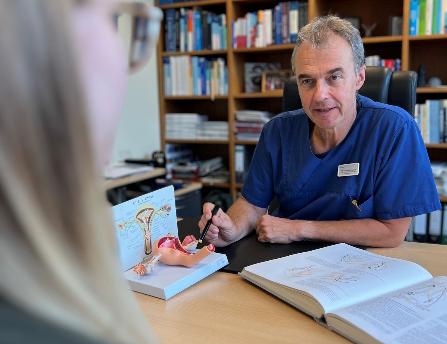
Lung cancer also remains the leading cause of cancer-related death globally, accounting for approximately 1.9 million deaths in 2024. One reason for its high mortality is that the disease is often diagnosed at an advanced stage: early symptoms such as fatigue or coughing can easily be mistaken for other conditions. In addition, some types of lung cancer tend to spread to other organs relatively early, making treatment more challenging. “At the same time, we know how important prevention, protection from tobacco smoke, and diagnosis at the earliest possible stage are. The earlier a tumor is detected, the better the chances of successful treatment,” says Prof. Dr. Tim Hirche, Director of the Department of Pulmonology at Helios Dr. Horst Schmidt Hospital Wiesbaden.
Causes and risk factors
The growth and division of cells in the body are normally tightly regulated: old cells die and are replaced by new ones. However, damage and genetic mutations can sometimes occur in a cell’s DNA. As a result of these changes, cells may begin to divide uncontrollably. When abnormal cells form a tumor that grows into surrounding tissues and can spread throughout the body, the disease is considered cancer.
The most important risk factor for lung cancer is smoking. Tobacco smoke contains thousands of chemicals, many of which are known to cause cancer or may contribute to its development. It is estimated that around 85% of lung cancer cases are associated with smoking.
Secondhand smoke also increases the risk. Regular exposure to tobacco smoke can raise the likelihood of developing lung cancer even in people who have never smoked themselves.
Other factors that may increase the risk of developing lung cancer include:
- air pollution, including exposure to fine particulate matter;
- occupational exposures, such as prolonged contact with asbestos, silica dust, and certain other carcinogenic substances;
- radon, a naturally occurring radioactive gas that can accumulate inside buildings, with concentrations varying considerably by region;
- hereditary predisposition and genetic factors, which may contribute to the development of lung cancer in some cases.
It is important to understand that lung cancer does not occur only in people who smoke, and having one or more risk factors does not mean that a person will necessarily develop the disease. At the same time, quitting smoking reduces the risk of lung cancer at any age. Stopping smoking can also be beneficial after a lung cancer diagnosis, as it may improve treatment outcomes and overall health.
Lung cancer diagnosis
Lung cancer diagnosis is not limited to detecting a tumor. It is also important to determine its exact location and size, identify the type of cancer cells, assess the lymph nodes, and establish whether the disease has spread to other organs. In non-small cell lung cancer, certain molecular characteristics of the tumor are also analyzed, as they may influence the choice of targeted therapy or immunotherapy.
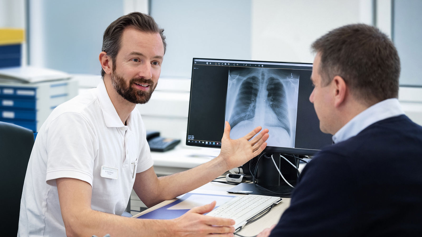
The diagnostic process usually begins with a medical consultation, review of the patient’s medical history, and assessment of symptoms. If a lung tumor is suspected, imaging tests are performed, followed in most cases by a biopsy to obtain a tissue sample and confirm the diagnosis. Once lung cancer has been confirmed, the stage of the disease is determined and an individual treatment plan is developed.
Early detection
Early-stage lung cancer often causes no noticeable symptoms. Cough, shortness of breath, fatigue, or weight loss can be nonspecific, which is one reason why the disease is sometimes diagnosed at a more advanced stage.
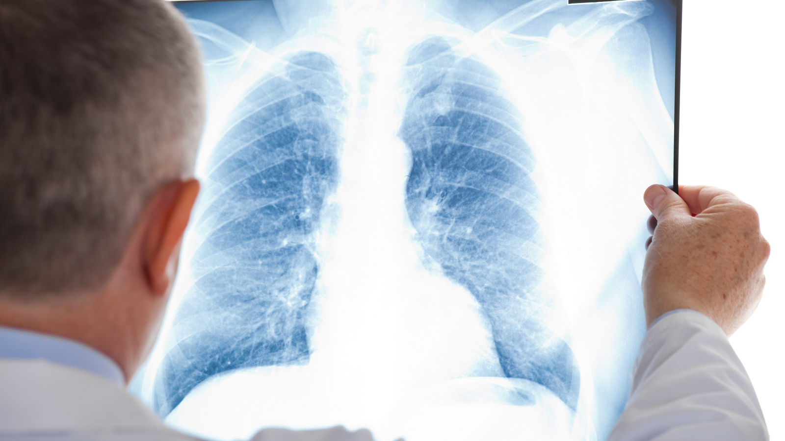
For people at high risk of developing lung cancer, the main method of early detection is low-dose computed tomography (LDCT). Screening is primarily intended for people with a long history of heavy smoking, including former smokers. Unlike a conventional chest X-ray, low-dose CT can detect small changes in the lungs at an earlier stage.
Eligibility criteria for lung cancer screening — including age, smoking history, and smoking intensity — vary between countries and according to national guidelines. The need for regular screening should therefore be discussed individually with a doctor.
Diagnostic methods
Several methods may be used to confirm the diagnosis and determine the extent of the disease:
- Computed tomography (CT) is one of the main imaging methods used when lung cancer is suspected. It helps determine the location, size, and characteristics of the tumor, assess the lymph nodes, and identify possible abnormalities in other organs.
- Bronchoscopy allows doctors to examine the airways from the inside using a thin, flexible endoscope and obtain tissue samples from suspicious areas for further analysis.
- Endobronchial ultrasound (EBUS) is performed during bronchoscopy and allows doctors to visualize and sample lymph nodes and other structures within the chest. It is particularly important for determining the stage of the disease.
- PET/CT combines computed tomography with information about the metabolic activity of tissues. It can help determine the extent of the cancer and identify possible metastases, for example before surgery or definitive radiation therapy.
- Biopsy and histopathological examination are used to confirm the diagnosis and determine the type of lung cancer. The tissue is examined under a microscope to distinguish, among other things, between non-small cell lung cancer (NSCLC) and small cell lung cancer (SCLC).
- Molecular diagnostics can identify specific genetic alterations and other tumor biomarkers. In non-small cell lung cancer, these findings may play a decisive role in selecting targeted therapy or immunotherapy.
Once the diagnosis has been confirmed, staging is performed to determine the size and local extent of the tumor, lymph node involvement, and the presence of distant metastases. Depending on the individual clinical situation, additional tests such as PET/CT, MRI, and other examinations may be required.
Ion robotic system for lung cancer diagnosis
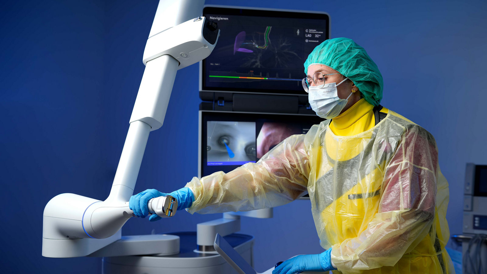
Small suspicious nodules located deep within the peripheral areas of the lungs can be particularly difficult to reach. With conventional bronchoscopy, accessing these lesions and obtaining enough tissue for analysis may be challenging. To address this, specialized Helios centers use the Ion robotic-assisted navigation bronchoscopy system.
Before the procedure, a high-resolution CT scan of the lungs is performed. Based on these images, specialized software creates a three-dimensional model of the airways and plans a route to the suspicious lesion. During bronchoscopy, the physician controls a very thin and flexible catheter that can be precisely guided through the branching airways toward small nodules in the peripheral areas of the lung.
Once the suspicious area has been reached, the physician can perform a targeted biopsy and obtain tissue samples for histopathological and molecular analysis. The Ion system can be particularly useful for small and difficult-to-reach lesions that may be challenging to access with conventional bronchoscopy.
Ion robotic-assisted bronchoscopy is used at specialized Helios lung cancer centers, including locations in Berlin, Wiesbaden, Bonn/Siegburg, Krefeld/Duisburg, and Erfurt. Precise navigation to small peripheral lung lesions can make it possible to obtain diagnostic tissue using a minimally invasive approach and provide the information needed to determine the next steps in treatment.
Read more about Ion robotic system ⟶
Lung cancer treatment
The choice of treatment for lung cancer depends on the type and stage of the tumor, its molecular characteristics, lung function, the patient’s overall health, and any underlying medical conditions. Treatment also differs considerably between the two main types of lung cancer: non-small cell lung cancer (NSCLC) and small cell lung cancer (SCLC).
“Today, we have significantly better treatment options for lung cancer than we did just a few years ago. Nevertheless, one principle remains unchanged: the earlier the tumor is detected, the better the chances of a cure.” — Priv.-Doz. Dr. med. Nikolaus Büchner, Head of the Department of Pulmonology, Sleep and Respiratory Medicine at Helios St. Johannes Hospital Duisburg.
For localized non-small cell lung cancer, treatment may aim to completely remove or destroy the tumor. Depending on the stage, this may involve surgery, radiation therapy, systemic therapy, or a combination of these approaches. Locally advanced disease often requires multimodal treatment, while systemic therapies generally play the central role in metastatic lung cancer.
For metastatic lung cancer, treatment is primarily aimed at controlling the disease, slowing its progression, relieving symptoms, and maintaining quality of life. With modern systemic treatments, long-term disease control may be possible for some patients.
Surgical treatment
Surgery is one of the main treatment options for early-stage non-small cell lung cancer and may offer the possibility of completely removing the tumor. During surgery, the surgeon removes the tumor together with the necessary amount of surrounding lung tissue and assesses or removes regional lymph nodes.
Depending on the size and location of the tumor, surgical procedures may include:
- Segmentectomy — removal of an anatomical segment of the lung.
- Lobectomy — removal of one lobe of the lung.
- Bilobectomy — removal of two lobes of the right lung.
- Pneumonectomy — removal of an entire lung.
The extent of surgery is selected with the aim of completely removing the tumor while preserving as much functioning lung tissue as possible. Before surgery, lung function and the patient’s ability to tolerate the planned procedure are carefully assessed.
For more complex, locally advanced tumors, specialized Helios thoracic surgery centers may also perform extended resections involving the bronchi, blood vessels, chest wall, diaphragm, or other adjacent structures when complete tumor removal is considered technically feasible.
Surgery is used much less frequently for small cell lung cancer and is generally considered only in selected patients with very early, localized disease.
Minimally invasive and robot-assisted surgery
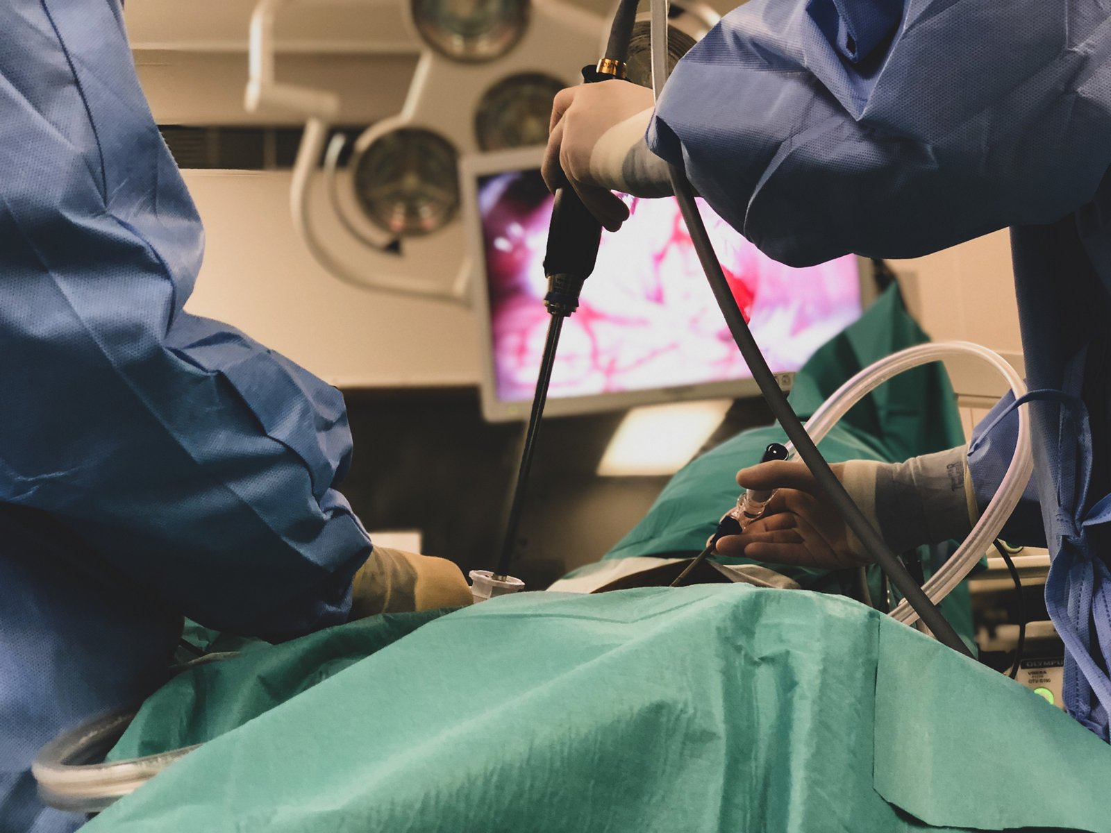
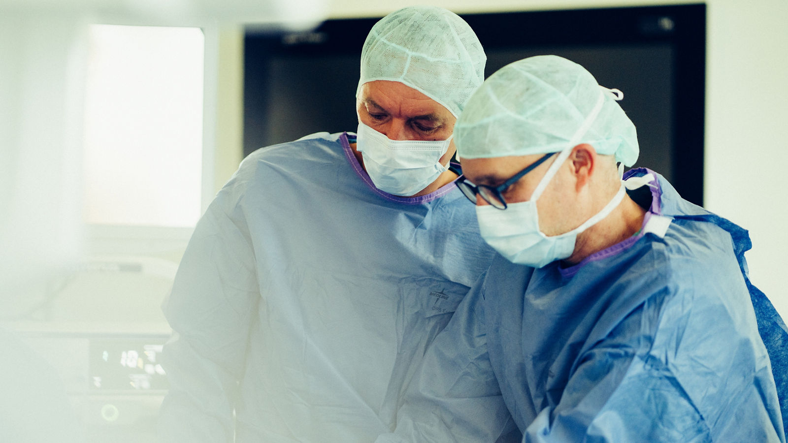
Many lung cancer operations can now be performed through small incisions using video-assisted thoracoscopic surgery (VATS). A camera and specialized surgical instruments are introduced through one or several small access points, allowing the surgeon to remove the affected part of the lung without widely opening the chest.
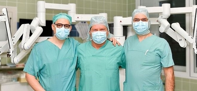
Some Helios centers also use robot-assisted thoracic surgery (RATS) with the Da Vinci surgical system. The surgeon controls the instruments from a console while viewing a magnified three-dimensional image of the surgical field. The high degree of instrument mobility enables precise surgical maneuvers within the confined space of the chest.
For example, robot-assisted lobectomies and segmentectomies are performed at Helios Hospital Bonn/Rhein-Sieg, while robotic thoracic surgery is also available at other specialized Helios centers.
A minimally invasive approach is not suitable for every patient. Large or locally advanced tumors may require open surgery.
Chemotherapy
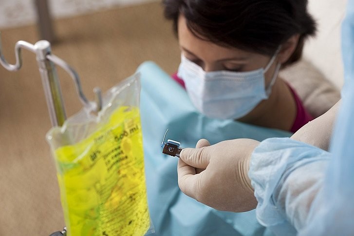
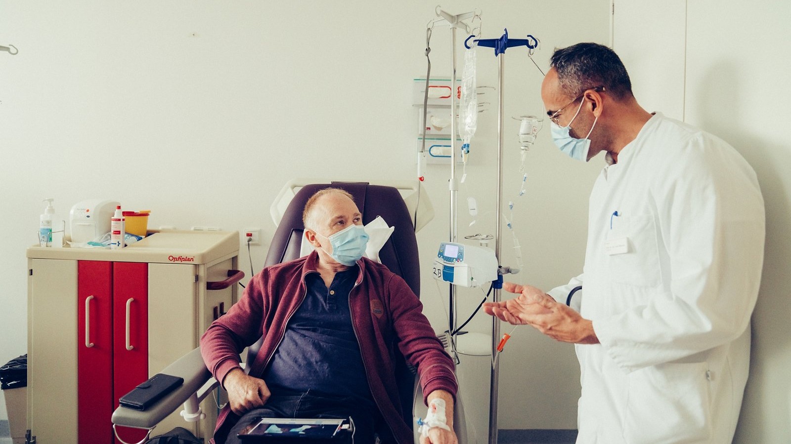
Chemotherapy uses anticancer drugs that target rapidly dividing cells throughout the body. It may be used on its own or in combination with surgery, radiation therapy, or immunotherapy.
Depending on the clinical situation, chemotherapy may be given:
- before surgery (neoadjuvant therapy) to reduce the tumor and treat possible microscopic cancer deposits;
- after surgery (adjuvant therapy) to destroy cancer cells that may remain in the body and reduce the risk of recurrence;
- together with radiation therapy, particularly for certain locally advanced tumors;
- for advanced or metastatic disease, either alone or in combination with immunotherapy.
Systemic drug therapy plays a particularly important role in small cell lung cancer because this type of cancer tends to spread throughout the body at an early stage.
Immunotherapy
Immunotherapy has become one of the major advances in modern lung cancer treatment. In particular, drugs known as immune checkpoint inhibitors are used in selected patients.
Cancer cells can exploit certain biological mechanisms to suppress the immune response and avoid being attacked by the immune system. Immunotherapy blocks these signals, helping the immune system recognize and attack cancer cells more effectively.
In non-small cell lung cancer, immunotherapy may be used at different stages of treatment: in combination with chemotherapy before surgery, as part of perioperative treatment before and after surgery, after surgery in selected patients, after chemoradiotherapy for certain locally advanced tumors, or in metastatic disease.
Whether immunotherapy is appropriate depends on the type and stage of the tumor, molecular findings, the expression of biomarkers such as PD-L1, and other clinical factors.
Targeted therapy
In some patients with non-small cell lung cancer, cancer cells contain specific genetic alterations that drive their growth. These are often referred to as driver mutations or actionable molecular targets.
If molecular testing identifies a suitable target, targeted drugs may be used to block the specific mechanism that promotes the growth and survival of the cancer cells. Well-established examples of clinically relevant alterations include EGFR mutations and ALK or ROS1 rearrangements, while modern comprehensive molecular testing can identify a number of additional actionable targets.
Targeted therapy is particularly important in advanced non-small cell lung cancer, although certain targeted treatments may also be used at earlier stages as part of a multimodal treatment strategy.
For this reason, molecular analysis of tumor tissue is an important part of modern NSCLC diagnostics. At Helios Hospital Emil von Behring in Berlin, molecular pathology testing is organized so that, in appropriate cases, results and treatment recommendations can be available within a few days.
Radiation therapy
Radiation therapy uses high-energy ionizing radiation to damage the DNA of cancer cells and destroy them. Modern treatment planning makes it possible to deliver radiation precisely to the tumor while limiting exposure to surrounding healthy tissues as much as possible.
The role of radiation therapy depends on the stage and clinical situation. It may be used:
- as a definitive treatment for early-stage lung cancer when surgery is not possible;
- together with chemotherapy for locally advanced disease;
- before or after other treatments as part of a multimodal strategy;
- to treat individual metastases, for example in the brain;
- as palliative treatment to relieve pain, bleeding, cough, shortness of breath, or other symptoms.
Stereotactic body radiation therapy (SBRT)
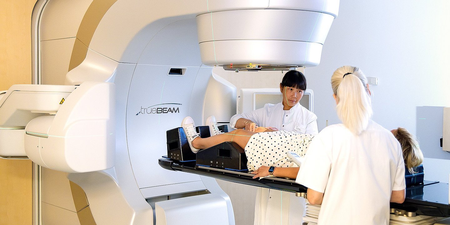
For small, localized tumors, stereotactic body radiation therapy (SBRT) may be used. This technique delivers a high dose of radiation very precisely to a defined target over a limited number of treatment sessions.
SBRT is particularly important for patients with early-stage lung cancer who cannot undergo surgery because of underlying medical conditions, reduced lung function, or other medical reasons. It may also be used to treat selected metastases.
Stereotactic radiation therapy for lung tumors is available at specialized Helios centers. Modern treatment-planning systems and linear accelerators allow radiation to be delivered with high precision while minimizing exposure to surrounding healthy tissue.
Interventional bronchoscopy
When a tumor grows within a major bronchus or compresses the airway, it can cause severe shortness of breath, coughing, coughing up blood, or impaired ventilation of part of the lung. In such situations, specialized Helios centers may use interventional bronchoscopy.
Using a bronchoscope, the physician can reach the tumor from inside the airways. Depending on the individual situation, treatment may include:
- mechanical removal or reduction of tumor tissue;
- laser therapy;
- cryotherapy or cryoresection, in which tumor tissue is treated using very low temperatures;
- argon plasma coagulation (APC);
- reopening of a narrowed bronchus;
- placement of a bronchial or tracheal stent to keep the airway open.
These procedures do not usually replace systemic cancer treatment, but they can rapidly restore airway patency, relieve symptoms, and make it possible to proceed with further anticancer therapy.
Local tumor ablation
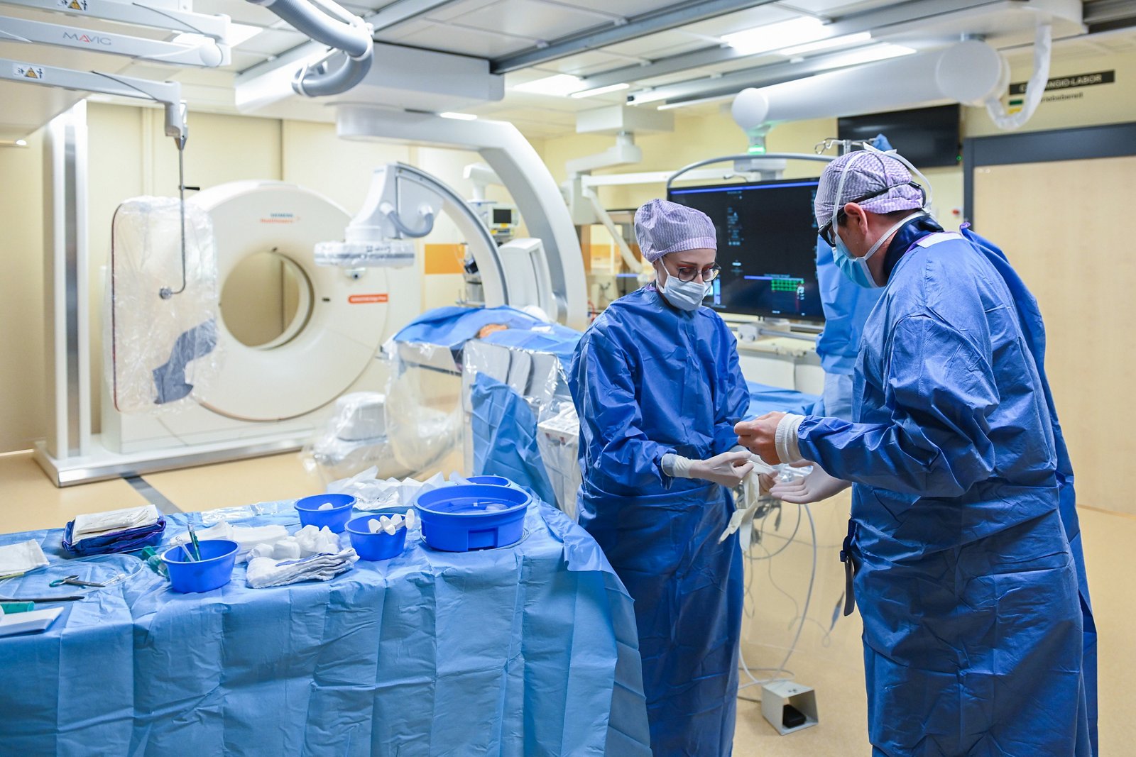
For selected patients with small lung tumors or a limited number of lesions, percutaneous tumor ablation may be considered. Under CT guidance, a physician introduces a special probe directly into the tumor through a small skin puncture and destroys the tumor tissue using energy.
Helios centers use techniques including radiofrequency ablation (RFA) and, at selected locations, microwave ablation (MWA) for lung tumors. These procedures are available, for example, through interventional radiology at Helios Hospital Krefeld and Helios Park Hospital Leipzig. In Leipzig, both RFA and MWA are offered for lung tumors using computer-assisted treatment planning.
Ablation is not a standard substitute for surgery for every patient with early-stage lung cancer. Its use is considered individually, for example when surgery would involve a high risk or when local control of a limited number of tumor lesions is required.
Follow-up care and rehabilitation
After completing lung cancer treatment, patients require regular medical follow-up. The aim is to monitor recovery, detect possible recurrence or metastases at an early stage, and identify and manage any long-term effects of surgery, radiation therapy, or systemic treatment.
The follow-up schedule is tailored to the type and stage of lung cancer, the treatment received, and the patient’s overall health. Follow-up usually includes medical consultations and computed tomography (CT) scans of the chest. Depending on the individual situation, additional tests may include laboratory examinations, lung function tests, bronchoscopy, PET/CT, MRI, or other diagnostic procedures. Follow-up is generally more frequent during the first years after treatment, with longer intervals between examinations later on.
After lung surgery, pulmonary and physical rehabilitation can be an important part of recovery. Breathing exercises, physiotherapy, and gradually increasing physical activity can help patients adapt to reduced lung capacity, improve breathing, and rebuild strength and endurance. The level of physical activity is tailored to the patient’s lung function and overall condition.
Rehabilitation may also include nutritional support, physiotherapy, pain and symptom management, and psycho-oncological support. Patients who experience persistent shortness of breath or a significant reduction in physical capacity may benefit from specialized pulmonary rehabilitation programs.
For patients who continue to smoke, smoking cessation is an important part of ongoing care. Quitting remains beneficial even after a lung cancer diagnosis: it supports the recovery of the respiratory and cardiovascular systems, reduces the risk of complications, and may improve treatment outcomes.
After active treatment has been completed, patients should also pay attention to new or worsening symptoms, such as a persistent cough, coughing up blood, increasing shortness of breath, unexplained weight loss, or new pain. If such symptoms occur, patients should contact their doctor rather than wait until the next scheduled follow-up appointment.
Individualized treatment and clinical trials at Helios
Modern lung cancer treatment is increasingly based not on a single method, but on an individually planned sequence of different therapies. The choice of treatment depends on the type and stage of the tumor, its molecular characteristics, lung function, the patient’s overall health, and any previous treatment. At specialized Helios centers, complex cases are reviewed by a multidisciplinary tumor board. Pulmonologists, thoracic surgeons, medical oncologists, radiation oncologists, radiologists, pathologists, and other specialists work together to review diagnostic findings and determine the most appropriate treatment strategy. When necessary, surgery can be combined with chemotherapy, immunotherapy, targeted therapy, or radiation therapy in a carefully planned sequence.
Depending on the individual clinical situation and the Helios center, patients may have access to minimally invasive and robot-assisted lung surgery, complex extended thoracic procedures, high-precision stereotactic radiation therapy, modern immunotherapies and targeted treatments, interventional bronchoscopy, and local tumor ablation. This broad range of treatment options can be particularly important in complex cases where achieving the best possible outcome requires expertise from several medical specialties.
Specialized centers can also provide a second medical opinion. If a patient has already received a diagnosis or treatment recommendation, Helios specialists can reassess imaging findings, histopathology and molecular test results, and discuss possible alternative treatment strategies.
Helios also participates in clinical trials investigating new drugs and treatment strategies for lung cancer. For selected patients, participation in a clinical trial may provide access to emerging therapies that are not yet part of standard treatment. Eligibility is assessed individually based on the type and stage of the tumor, previous treatment, and the specific criteria of each study.
For international patients, treatment can be coordinated through Helios International, from the initial review of medical records and selection of an appropriate hospital and specialist to the organization of diagnostics, treatment, and follow-up care. This can be particularly valuable in lung cancer, where treatment decisions often require input from several specialists as well as timely additional testing, including molecular diagnostics.
The goal at Helios is therefore not simply to select an individual treatment method, but to develop a personalized treatment strategy that takes into account the biological characteristics of the tumor, the extent of the disease, and the patient’s overall condition, bringing the necessary diagnostic and therapeutic approaches together in a coordinated treatment plan.
Learn more about arranging treatment at Helios hospitals ⟶
Read more:
Do you need more information about Helios Hospitals or want to schedule your treatment?