Testicular cancer treatment: Modern methods and treatment options
- Causes and risk factors
- Testicular cancer diagnosis
- Testicular cancer treatment
- Follow-up care and rehabilitation
- Fertility after testicular cancer
- Specialized testicular cancer treatment at Helios
The testicles are paired organs of the male reproductive system located in the scrotum. They have two main functions: producing sperm and male sex hormones, primarily testosterone.
Testicular cancer develops when cells in the testicular tissue begin to divide uncontrollably and form a malignant tumor. In most cases, the disease originates from germ cells — the cells involved in sperm production. These tumors are known as germ cell tumors and are divided into two main groups: seminomas and non-seminomatous germ cell tumors. This distinction is important because these tumor types can behave differently and may require different approaches to treatment.
Unlike many other cancers, testicular cancer occurs most frequently in younger men, and in most cases only one testicle is affected. At an early stage, the disease often causes no pain and may first become noticeable as a painless lump, enlargement of the testicle, or a change in its shape or consistency.
“Testicular cancer is most commonly diagnosed in men between the ages of 20 and 45. Overall, the disease primarily affects men between the ages of 15 and 45.” — Dr. med. Inga Kunz, Senior Consultant and Site Lead at the Department of Urology and Pediatric Urology at Helios St. Josefshospital Uerdingen.
At the same time, testicular cancer is considered one of the most curable cancers. Modern treatments can achieve a cure in the vast majority of patients, and successful treatment is often possible even when the disease has spread beyond the testicle. The earlier the tumor is detected, the less intensive the subsequent treatment may need to be.
Causes and risk factors
The exact causes of testicular cancer are not fully understood. The disease develops as a result of changes in the cells of the testicular tissue that cause them to grow uncontrollably and form a tumor. However, several factors are known to increase the risk of developing testicular cancer.
The main risk factors include:
- Undescended testicle (cryptorchidism). During fetal development, the testicles form in the abdomen and gradually descend into the scrotum. If one or both testicles do not descend completely, the risk of developing testicular cancer later in life is increased. Some increased risk remains even after surgical correction of cryptorchidism during childhood.
- Family history. The risk is higher in men who have a close relative with testicular cancer, particularly a father or brother. This suggests that inherited factors may contribute to the development of some testicular tumors.
- Previous testicular cancer. Men who have previously had cancer in one testicle have a higher risk of developing cancer in the other testicle compared with men who have never had the disease.
- Certain genetic factors. Some genetic and chromosomal abnormalities may be associated with an increased risk of developing testicular tumors.
Having one or more risk factors does not mean that a person will necessarily develop testicular cancer. Conversely, the disease can occur in men who have no known risk factors.
Because testicular cancer most commonly affects younger men and often causes no pain in its early stages, it is important to pay attention to changes in the testicles. A painless lump, enlargement of a testicle, or a change in its shape or consistency should be evaluated by a urologist.
Testicular cancer diagnosis
Testicular cancer is often first noticed by the patient, for example as a painless lump, enlargement of a testicle, or a change in its shape or consistency. If such changes occur, it is important to see a urologist. Most abnormalities in the scrotum are not cancer, but their cause can only be determined through medical evaluation.
Diagnosis is aimed not only at confirming the presence of a tumor but also at determining its type and whether the disease has spread. This information is essential for selecting the appropriate treatment.
Testicular self-examination
Being familiar with how the testicles normally look and feel can make it easier to notice unusual changes. Self-examination can be performed after a warm shower or bath, when the skin of the scrotum is relaxed.
Each testicle can be gently rolled between the thumb and fingers, checking for new lumps, enlargement or shrinkage, changes in shape or consistency, or a feeling of heaviness in the scrotum. A slight difference in the size or position of the two testicles can be normal.
A soft structure can also be felt behind each testicle. This is the epididymis, a normal part of the male reproductive system that can sometimes be mistaken for an abnormal lump.
Self-examination does not replace an examination by a urologist and cannot be used to diagnose testicular cancer. A new lump, swelling, noticeable change in size, or any other unusual change should be evaluated by a doctor, even if it is painless.
Medical diagnosis
The diagnostic process usually begins with a consultation with a urologist. The doctor will ask about symptoms, how long the changes have been present, previous medical conditions, and possible risk factors, followed by a physical examination of the testicles and scrotum.
The main diagnostic methods include:
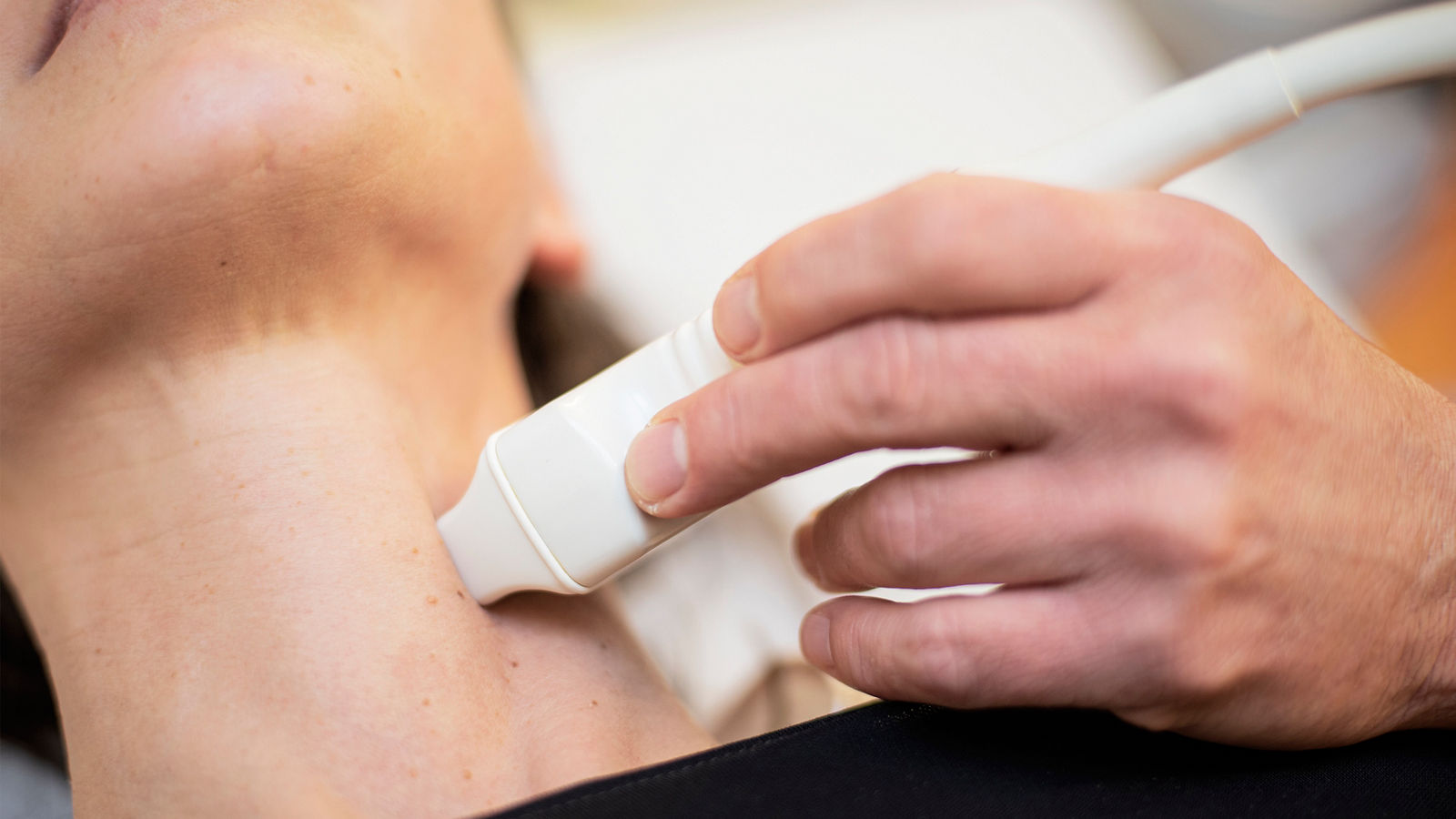
- Testicular ultrasound. This is the primary imaging method used when a testicular tumor is suspected. Ultrasound helps determine the location, size, and characteristics of a mass and distinguish abnormalities within the testicular tissue from those located outside the testicle.
- Blood tests for tumor markers. When testicular cancer is suspected, the main markers measured are AFP (alpha-fetoprotein), β-hCG (beta-human chorionic gonadotropin), and LDH (lactate dehydrogenase). These markers can help with diagnosis, disease classification, prognosis, and subsequent monitoring of treatment response. However, normal tumor marker levels do not rule out testicular cancer.
- Computed tomography (CT). If the diagnosis has been confirmed or there is a strong suspicion of malignancy, CT scans of the chest, abdomen, and pelvis may be used to assess lymph nodes and look for possible metastases.
- Magnetic resonance imaging (MRI). MRI may be used in selected situations, for example when other imaging findings are inconclusive or when additional evaluation of specific areas is required.
When a malignant testicular tumor is suspected, a conventional needle or transscrotal biopsy is generally avoided. If the findings indicate cancer, the standard next step is usually surgical removal of the affected testicle through an incision in the groin — a procedure known as radical inguinal orchiectomy. The removed tissue is then examined in the laboratory to confirm the diagnosis and determine the exact type of tumor.
Once the diagnosis has been confirmed, staging is performed. Doctors assess the characteristics of the primary tumor, lymph node involvement, the presence of distant metastases, and tumor marker levels. Based on these findings, the stage of the disease is determined and the next treatment strategy is selected, which may include active surveillance, chemotherapy, radiation therapy, or additional surgery.
Testicular cancer treatment
Testicular cancer is one of the cancers with a very high likelihood of successful treatment. The choice of therapy depends primarily on the type of tumor — seminoma or non-seminomatous germ cell tumor — as well as the stage of the disease, tumor marker levels, and the presence of metastases.
Treatment may include surgery, active surveillance, chemotherapy, radiation therapy, or a combination of several approaches. Even when the disease has spread beyond the testicle, treatment can often still be given with the goal of achieving a complete cure.
Surgical removal of the testicle
When a malignant testicular tumor is suspected, the first step in treatment is usually a radical inguinal orchiectomy — removal of the affected testicle through a small incision in the groin.
The removed tissue is examined in the laboratory. Histopathological analysis determines the exact type of tumor, in particular whether it is a seminoma or a non-seminomatous germ cell tumor. Together with imaging findings and tumor marker levels, this information is used to determine the stage of the disease and plan further treatment.
For some patients, surgery is the only cancer treatment required. If the disease is confined to the testicle and the risk of recurrence is low, no additional treatment may be necessary after surgery. In other cases, depending on the type and stage of the tumor, chemotherapy, radiation therapy, or additional surgery may be recommended.
Removal of one testicle usually does not cause a significant decrease in testosterone levels or affect sexual function if the remaining testicle is functioning normally. A testicular prosthesis can be inserted if the patient wishes.
In selected situations, such as a tumor in a solitary testicle or certain bilateral tumors, testis-sparing surgery may be considered. The suitability of this approach is assessed individually.
Chemotherapy
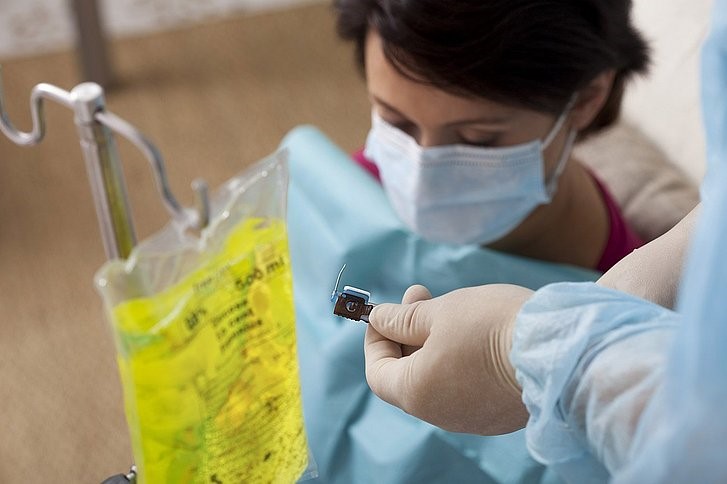
Chemotherapy is used primarily for advanced testicular cancer, when cancer cells have spread to lymph nodes or other organs. It may also be recommended after removal of the testicle for some patients who have a higher risk of recurrence.
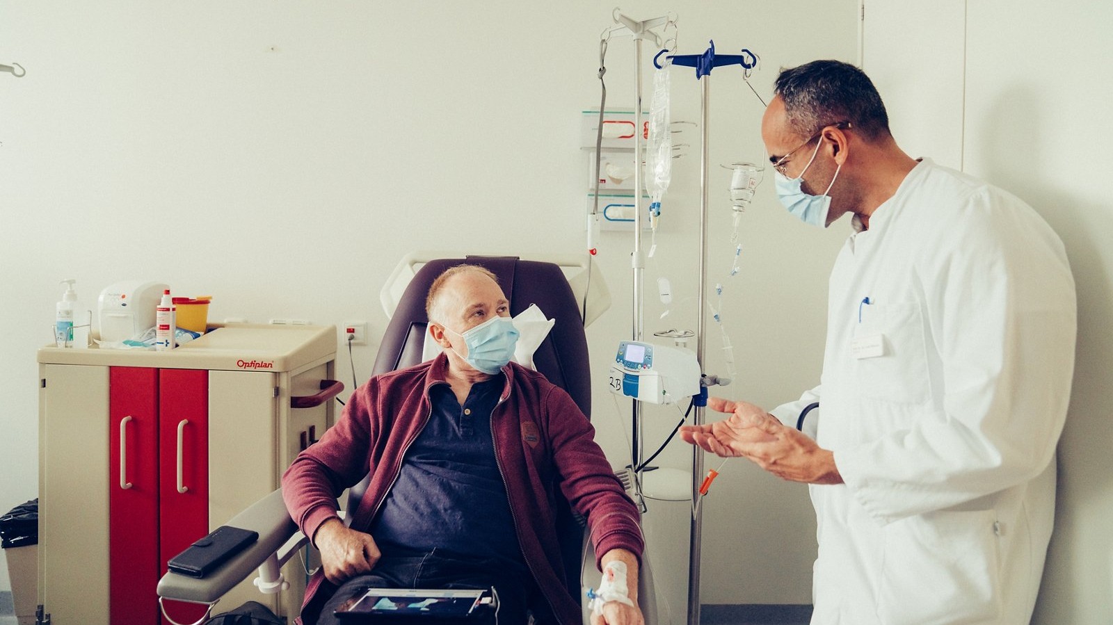
Testicular germ cell tumors are highly sensitive to platinum-based chemotherapy. One of the standard combinations is BEP, which consists of bleomycin, etoposide, and cisplatin. Other regimens may be used in certain situations. The choice of drugs and number of treatment cycles depend on the type of tumor, stage of the disease, and prognostic group.
For metastatic non-seminomatous germ cell tumors, systemic chemotherapy is one of the main treatment approaches. It is also widely used for advanced seminoma.
Specialized chemotherapy for testicular cancer is available, among other centers, at the Testicular Cancer Center at Helios Hospital Bad Saarow. The center has many years of experience in both systemic and surgical treatment of malignant testicular tumors. Treatment is provided within an interdisciplinary cancer center.
Radiation therapy
The role of radiation therapy differs considerably depending on the type of tumor.
Seminomas are highly sensitive to radiation, so radiation therapy may be used in selected patients, particularly in certain cases involving regional lymph nodes.
Today, this treatment is used more selectively than in the past. Because testicular cancer often affects young men who have a high likelihood of long-term survival, the potential long-term effects of radiation need to be taken into account when choosing treatment.
Non-seminomatous germ cell tumors do not have the same sensitivity to radiation, so radiation therapy is generally not a standard treatment for these tumors.
Retroperitoneal lymph node dissection
One of the typical routes of testicular cancer spread is to the retroperitoneal lymph nodes, which are located deep in the abdomen near major blood vessels. In certain situations, particularly in patients with non-seminomatous germ cell tumors, these lymph nodes may need to be surgically removed. This procedure is known as retroperitoneal lymph node dissection (RPLND).
RPLND may be used in selected patients with involvement of the retroperitoneal lymph nodes, as well as after chemotherapy if residual tumor masses remain.
Whenever possible, a nerve-sparing technique is used. Surgeons aim to preserve the sympathetic nerve fibers involved in ejaculation, which is particularly important when treating young patients.
Nerve-sparing RPLND for testicular cancer is performed, among other centers, at Helios Dr. Horst Schmidt Hospital Wiesbaden and Helios University Hospital Wuppertal.
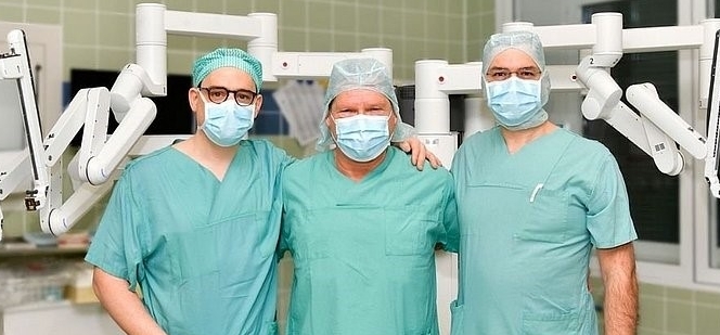
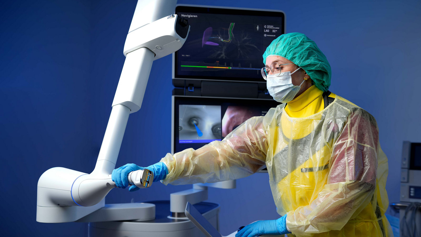
At some Helios centers, RPLND can also be performed using Da Vinci robot-assisted surgery. This approach is available, for example, at Helios Hospital Bad Saarow and Helios Hospital Berlin-Buch. The robotic system provides the surgeon with a magnified three-dimensional view of the surgical field and enables highly precise instrument movements when operating in the anatomically complex area surrounding major blood vessels.
The choice between open and robot-assisted surgery depends on the location and size of the tumor masses, previous treatment, and the individual clinical situation.
Removal of residual tumors after chemotherapy
After chemotherapy, metastases may shrink considerably. However, follow-up imaging may still show residual tumor masses in some patients. Their evaluation is particularly important in non-seminomatous germ cell tumors. A residual mass may consist of scar tissue, mature teratoma, or viable cancer cells. Surgical removal may therefore be necessary in certain situations.
These procedures can be considerably more complex than the initial surgery to remove the testicle. Residual tumors are often located in the retroperitoneum directly adjacent to the aorta, inferior vena cava, and other important structures, and may sometimes involve surrounding tissues.
Helios Hospital Berlin-Buch specializes in complex surgery to remove residual tumors in patients with testicular cancer. In suitable cases, these procedures can also be performed with the Da Vinci Xi robotic system.
Surgical removal of residual tumors after systemic chemotherapy is also part of the specialized treatment available at Helios centers in Bad Saarow and Wiesbaden.
Follow-up care and rehabilitation
After treatment for testicular cancer, patients require regular medical follow-up. The main goals are to detect a possible recurrence at an early stage, monitor recovery, and identify any potential long-term effects of treatment.
The follow-up schedule is tailored to each patient and depends on the type and stage of the tumor, the treatment received, and the risk of recurrence. In general, follow-up appointments are more frequent during the first few years, when the risk of the cancer returning is highest.
Follow-up examinations
Follow-up care may include:
-
medical consultations and physical examinations;
-
blood tests for the tumor markers AFP, β-hCG, and LDH;
-
computed tomography (CT) or other imaging tests depending on the stage of the disease and previous treatment;
-
monitoring of testosterone and other hormone levels if relevant symptoms occur.
The frequency and type of examinations gradually change over time. It is important to follow the recommended schedule even when feeling well, as an early recurrence may not cause noticeable symptoms.
For patients with early-stage disease who do not require additional treatment after removal of the testicle, regular follow-up examinations are part of an active surveillance strategy. This approach can help avoid unnecessary chemotherapy or radiation therapy while ensuring that treatment can be started promptly if signs of recurrence appear.
Recovery after treatment
After removal of one testicle, most patients can gradually return to their usual level of physical activity. The recovery period depends on the extent of surgery and any additional treatment received.
Recovery may take longer after chemotherapy. Fatigue, reduced physical stamina, and other side effects may persist for some time. Gradually increasing physical activity, maintaining a balanced diet, and getting adequate rest can support recovery.
Patients who have undergone intensive or combined treatment may, when needed, benefit from specialized cancer rehabilitation. This can include physical therapy, psychological support, and assistance with returning to everyday activities and work.
Fertility after testicular cancer
Preserving the ability to have children is an important consideration in testicular cancer, as the disease often affects younger men. For many patients, fertility is preserved after removal of one testicle if the remaining testicle functions normally. However, sperm production can be affected not only by treatment but also by the disease itself.
Chemotherapy and radiation therapy can temporarily or, less commonly, permanently reduce sperm production. Certain operations on the retroperitoneal lymph nodes can also affect ejaculation. For this reason, reproductive health and future family planning should ideally be discussed before treatment begins.
Men who may wish to have children in the future can be offered sperm cryopreservation before chemotherapy, radiation therapy, or other treatments that may affect reproductive function. Sperm is collected, frozen, and stored for possible future use with assisted reproductive technologies.
During retroperitoneal lymph node dissection, specialized Helios centers use nerve-sparing surgical techniques whenever possible to preserve the nerve fibers involved in ejaculation.
After treatment, a semen analysis can be performed when appropriate to assess the recovery of reproductive function. The rate of recovery varies from person to person and depends on fertility before treatment, the type and intensity of therapy received, and other individual factors.
Hormonal health and sexual function
If the remaining testicle functions normally, it can usually produce enough testosterone after removal of one testicle, so most patients do not require testosterone replacement therapy. If persistent fatigue, reduced libido, erectile dysfunction, loss of muscle mass, or other possible signs of testosterone deficiency occur, hormone levels can be checked.
If both testicles have been removed, the body can no longer produce sufficient testosterone on its own, and long-term testosterone replacement therapy is required.
Changes in appearance after surgery can also affect body image and sexual well-being. If the patient wishes, a testicular prosthesis can be inserted after removal of the testicle to recreate the appearance and feel of a testicle within the scrotum.
Specialized testicular cancer treatment at Helios
At Helios hospitals, testicular cancer treatment is planned by multidisciplinary teams involving urologists, oncologists, radiation oncologists, radiologists, and pathologists. Patients have access to modern diagnostic and treatment options, ranging from organ-preserving and nerve-sparing procedures to robot-assisted surgery, systemic therapies, and complex surgery following chemotherapy.
Particular attention is given to choosing the appropriate extent of treatment. Because testicular cancer has a high likelihood of cure, the goal is not only to treat the disease effectively but also to minimize long-term side effects and preserve hormonal function, fertility, and quality of life whenever possible.
Helios also provides expert second opinions, including for patients with complex or previously treated tumors. For international patients, Helios International coordinates the preliminary review of medical records, selection of an appropriate specialist, and the different stages of diagnosis and treatment.
Learn more about arranging treatment at Helios hospitals ⟶
Do you need more information about Helios Hospitals or want to schedule your treatment?