
Colorectal cancer treatment
- Symptoms of colorectal cancer
- Types of colorectal cancer
- Treatment methods depending on the stage of the disease
- Surgical treatment of colorectal cancer
- Chemotherapy for colorectal cancer
- Radiation therapy for colorectal cancer
- Targeted therapy for colorectal cancer
- Radionuclide therapy with lutetium-177
- Immunotherapy for colorectal cancer
- Follow-up care and rehabilitation
- Colorectal cancer treatment at Helios hospitals
Colorectal cancer is common in both men and women. According to global statistics, it is the third most commonly diagnosed cancer in men, after lung and prostate cancer. Among women, colorectal cancer also ranks third, following breast and lung cancer.
Important: Although colorectal cancer predominantly affects older adults, with the average age at diagnosis being approximately 71–75 years, the incidence among younger patients under the age of 50 has increased particularly sharply in recent years.
Symptoms of colorectal cancer
The early signs of colorectal cancer are often non-specific. Possible symptoms may include cramping abdominal pain or changes in bowel habits, such as constipation and diarrhea. However, similar symptoms can also occur with other conditions, including inflammatory bowel diseases such as Crohn’s disease and ulcerative colitis, or hemorrhoids. For this reason, they are not always immediately associated with a tumor. If symptoms persist for an extended period, it is advisable to consult a doctor and undergo appropriate examination.
Some of the most common possible signs include:
- changes in bowel habits — constipation and/or diarrhea, including alternating between the two;
- visible or occult blood in the stool;
- so-called “pencil-thin stools” — unusually narrow stools;
- mucus in the stool;
- unusual bowel sounds and bloating, sometimes accompanied by involuntary stool leakage;
- abdominal cramps and pain, as well as pain during bowel movements;
- palpable lumps or areas of hardness in the abdomen;
- unexplained weight loss;
- muscle weakness;
- fatigue and reduced physical performance;
- paleness;
- rapid heartbeat or increased heart rate even during mild physical activity.
Important: Intestinal polyps are among the most important precursors of colorectal cancer: approximately 90% of colorectal carcinomas develop from polyps that are initially benign.
Types of colorectal cancer
In general, colorectal cancer is divided into colon cancer (colon carcinoma) and rectal cancer (rectal carcinoma). Colon cancer accounts for approximately two-thirds of all colorectal cancer cases.
Depending on the location of the tumor, colon cancers can be classified as:
-
tumors of the cecum and ascending colon (colon ascendens);
-
tumors of the right colonic flexure (hepatic flexure) and the first part of the transverse colon (proximal transverse colon);
-
tumors of the middle part of the transverse colon;
-
tumors of the distal transverse colon and the left colonic flexure (splenic flexure);
-
tumors of the descending colon (colon descendens) and the proximal part of the sigmoid colon;
-
tumors of the middle and distal parts of the sigmoid colon.
Treatment methods depending on the stage of the disease
The choice of treatment for colorectal cancer depends on both the stage of the disease and the location of the tumor. Below are the main treatment approaches according to the stage of colorectal cancer.
Stage 0 (“Carcinoma in Situ”)
This is a very early stage of the disease in which the tumor is limited to the superficial layer of the lining of the colon. In many cases, endoscopic removal of the tumor is sufficient.
Stage I
The tumor has grown deeper into the intestinal wall and may involve the muscle layer, but the lymph nodes are not affected and there are no distant metastases. Some tumors at this stage can be completely removed endoscopically.
If certain risk factors are present, surgical removal of the affected section of the bowel may be necessary. Chemotherapy or radiation therapy is generally not required. Regular follow-up examinations are recommended after treatment to detect a possible recurrence at an early stage.
Stage II
The tumor has grown through the wall of the colon or rectum and may have spread into surrounding tissues, but the regional lymph nodes are not affected and there are no distant metastases. For colon cancer, the main treatment at this stage is usually surgical removal of the tumor.
The approach may be different for rectal cancer. Depending on the location and characteristics of the tumor, combined treatment with radiation therapy and chemotherapy may be given before surgery. The aim is to shrink the tumor and reduce the risk of recurrence. Certain risk factors and the molecular and genetic characteristics of the tumor may also influence the decision to use additional systemic therapy.
Stage III
At this stage, cancer cells have spread to the regional lymph nodes, but there are no distant metastases in other organs. For colon cancer, treatment usually involves surgical removal of the tumor and the affected section of the bowel, followed by adjuvant chemotherapy.
For rectal cancer, preoperative radiation therapy and chemotherapy or total neoadjuvant therapy are often used before surgery. After surgery, the further treatment strategy is determined individually, usually by a multidisciplinary tumor board.
Stage IV
In stage IV colorectal cancer, distant metastases are present, most commonly in the liver or lungs. Treatment is determined by a multidisciplinary team and depends on the extent of the disease, the location and number of metastases, the molecular characteristics of the tumor, and the patient’s overall health.
If both the primary tumor and the metastases can be completely removed, surgery may be performed on the bowel as well as on the organs affected by metastases. In other cases, systemic drug therapy is the main treatment approach and may include chemotherapy, targeted therapy, and, for tumors with certain molecular characteristics, immunotherapy. In some cases, systemic treatment can shrink metastases sufficiently to make previously inoperable lesions suitable for surgical removal. Surgery may also be necessary in cases of bowel obstruction or other complications caused by the primary tumor.
Surgical treatment of colorectal cancer
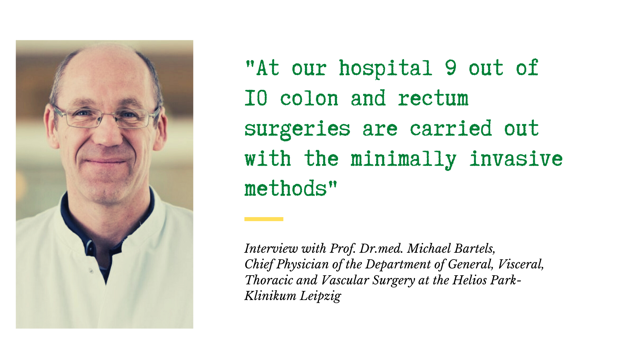
Surgery is one of the main treatment methods for colorectal cancer. Procedures can be performed using conventional open surgery or minimally invasive techniques, including laparoscopic and robot-assisted surgery.
The choice of surgical approach depends on a number of individual factors. These include the extent of the disease, the patient’s age and overall health, underlying medical conditions and previous surgeries, as well as the experience of the surgeon and surgical team and the patient’s preferences. The most appropriate surgical approach is determined in consultation with the treating physicians.
Minimally invasive surgery
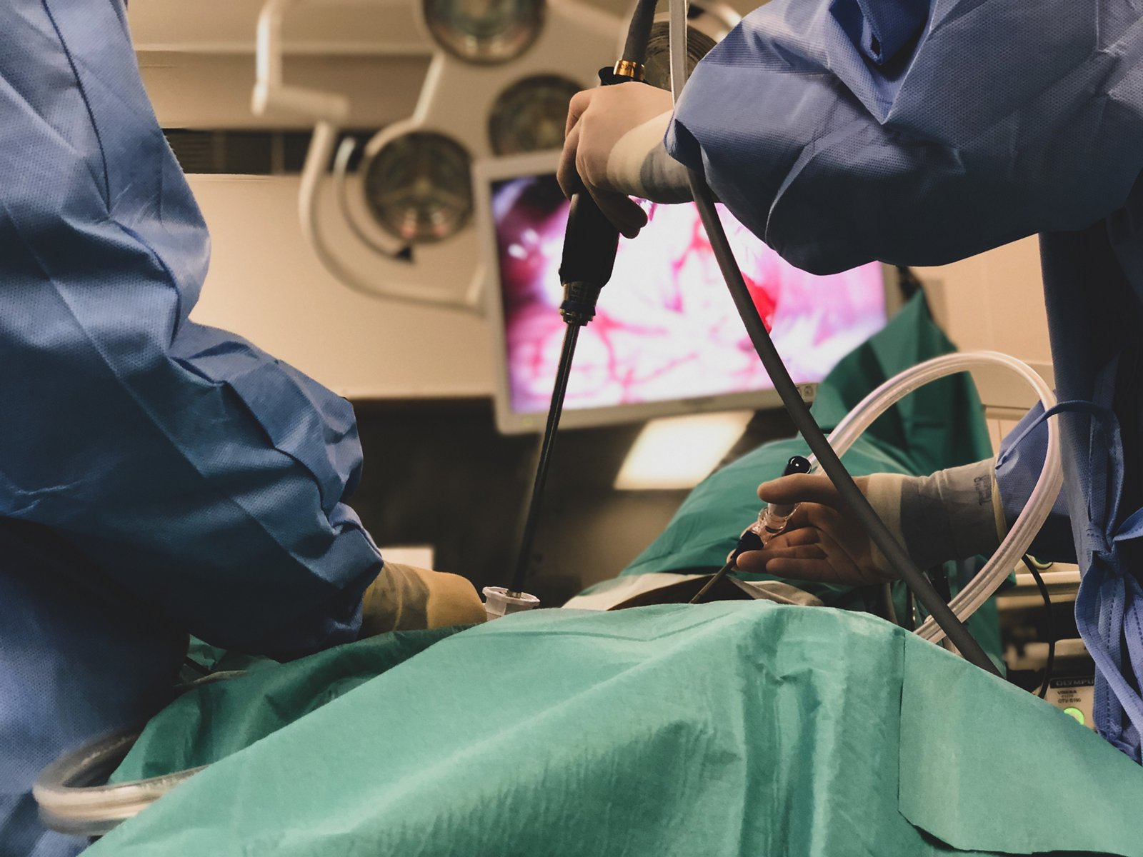
During minimally invasive surgery, the surgeon makes several small incisions, usually around 5 to 12 mm, through which specialized surgical instruments and a camera are inserted into the abdominal cavity. The camera provides a detailed view of the surgical field, allowing the surgeon to perform the necessary procedures, including removal of the tumor and the affected section of the bowel.
To create sufficient working space and improve visibility during the procedure, the abdominal cavity is inflated with carbon dioxide (CO₂). This lifts the abdominal wall and provides the surgeon with the space needed to perform the operation. The gas is released at the end of the procedure.
Robot-assisted surgery for colorectal cancer
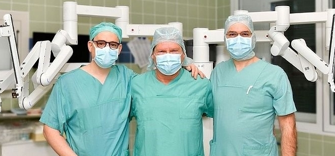
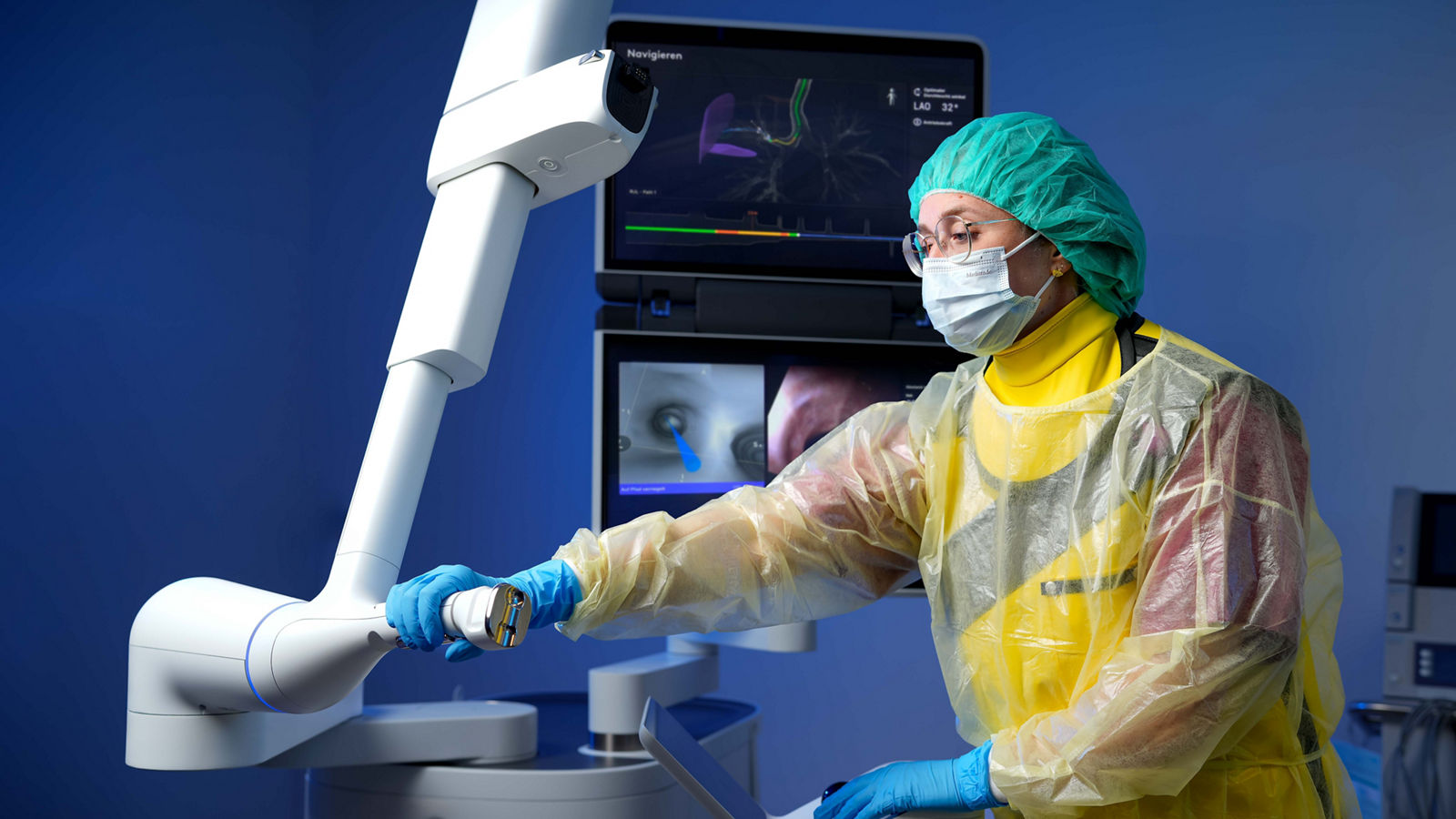
Robot-assisted surgery is a modern form of minimally invasive treatment for colorectal cancer. The procedure is performed through several small incisions using the da Vinci robotic surgical system, which is controlled by the surgeon from a console. The system provides a magnified, three-dimensional view of the surgical field and enables highly precise control of miniature surgical instruments. It filters out natural hand tremor, while the instruments’ wide range of motion gives the surgeon greater freedom of movement compared with conventional laparoscopy.
These capabilities are particularly valuable in rectal cancer surgery, where the surgeon must operate within the confined space of the pelvis, remove the tumor while maintaining oncological principles, and preserve surrounding nerves and other important anatomical structures as much as possible. Robot-assisted techniques are also used in colon cancer surgery. Small incisions and reduced tissue trauma may contribute to less blood loss and postoperative pain, faster recovery of bowel function, and earlier mobilization.
“The more complex the surgical procedure, the more the da Vinci system can demonstrate its capabilities and advantages.”
— Dr. Markus Mille, Senior Consultant in General and Visceral Surgery at Helios Hospital Erfurt.
Read more about robotic surgery for colon cancer using the da Vinci system ⟶
Artificial bowel opening (Stoma)
Today, most tumors of the colon and rectum can be surgically removed while preserving the natural route of bowel evacuation. However, in some cases, the treatment of colorectal cancer requires the creation of an artificial bowel opening, known as a stoma, through which intestinal contents are diverted to an opening in the abdominal wall and collected in a special stoma bag.
The need for a stoma depends primarily on the location and extent of the tumor, the type of surgery, and the condition of the bowel. For example, a permanent stoma may be necessary if a rectal tumor is located very close to the anal sphincter and complete removal of the tumor requires removal of the sphincter as well. The decision may also depend on sphincter function, the patient’s overall health, underlying medical conditions, and other individual factors.
However, a stoma does not always have to be permanent. In many cases, the surgeon creates a temporary stoma to divert intestinal contents away from the area where the ends of the bowel have been reconnected after tumor removal. This reduces stress on the intestinal anastomosis and allows it to heal properly. This approach is particularly common after rectal cancer surgery.
Once the bowel and the surgical connection have healed sufficiently, the physician assesses whether the stoma can be reversed and the natural passage of intestinal contents restored. This requires another operation in which bowel continuity is restored and the opening in the abdominal wall is closed. The timing of stoma reversal varies from patient to patient and depends on the healing process, subsequent cancer treatment, and the patient’s overall condition. If restoring the natural bowel passage is not possible or considered unsafe, the stoma may remain permanent.
Chemotherapy for colorectal cancer
In the treatment of colorectal cancer, chemotherapy may be administered before surgery or after the surgical procedure.
Chemotherapy given before surgery is known as neoadjuvant chemotherapy. Its aim is to shrink the tumor and create more favorable conditions for surgical removal. Adjuvant chemotherapy, on the other hand, is administered after surgery and is intended to reduce the risk of cancer recurrence.
If a multidisciplinary tumor board recommends adjuvant chemotherapy, treatment is generally started at an appropriate time after surgery, taking the patient’s recovery into account. The duration of chemotherapy depends on the stage of the disease and the treatment regimen selected and may, for example, last three or six months.
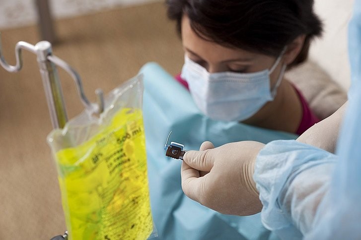
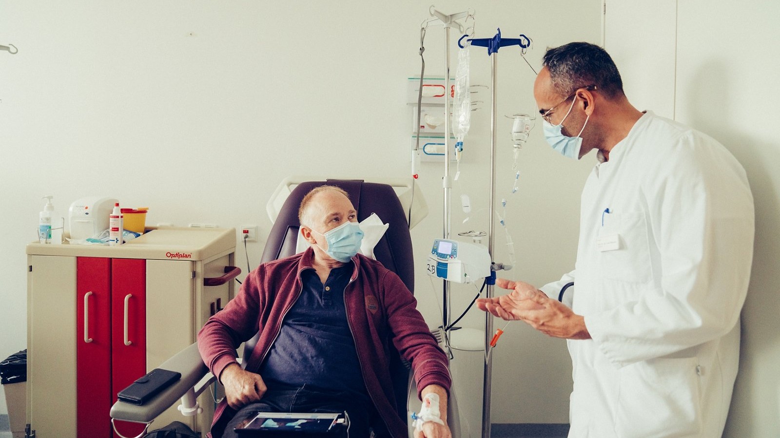
Depending on the medication used, chemotherapy may be administered orally in tablet form or intravenously by infusion. If long-term intravenous treatment is required, a port catheter may be implanted to provide convenient and reliable venous access.
Chemotherapy uses cytotoxic drugs that primarily target rapidly dividing cells, preventing them from multiplying and ultimately causing cell death. Because cytotoxic drugs can affect not only cancer cells but also some healthy rapidly dividing cells, treatment may be associated with various side effects.
For colon cancer, adjuvant chemotherapy is a standard part of treatment for stage III disease and may also be recommended for selected patients with stage II cancer who have high-risk features. For stage I colon cancer, chemotherapy is generally not required after complete surgical removal of the tumor.
For rectal cancer, the treatment sequence may differ. Depending on the stage, location, and characteristics of the tumor, systemic therapy may be given before surgery, including as part of total neoadjuvant therapy (TNT), sometimes in combination with radiation therapy. The optimal treatment strategy is determined individually by a multidisciplinary team.
Whether chemotherapy can be administered also depends on the patient’s overall health and any underlying medical conditions. Severe heart disease, significant impairment of liver or kidney function, and other serious conditions may influence the choice of medications, their dosage, or whether chemotherapy can be given at all.
Read more about chemotherapy for colorectal cancer ⟶
Radiation therapy for colorectal cancer
Radiation therapy may be administered either before or after surgery. Unlike chemotherapy, radiation therapy is a local treatment: its effects are directed at cancer cells within the irradiated area.
Radiation therapy given before surgery is known as neoadjuvant radiation therapy. Its aim is to shrink the tumor and create more favorable conditions for surgery. In some cases, a complete clinical response may be achieved. According to current evidence, selected patients with rectal cancer who achieve a complete clinical response after neoadjuvant chemoradiotherapy may be considered for a “Watch and Wait” strategy. With careful and regular monitoring, this approach may allow some patients to avoid immediate surgery and preserve the rectum. The decision is made individually at a specialized center.
Adjuvant radiation therapy is administered after surgical removal of the tumor and may be used to reduce the risk of local recurrence. The need for radiation therapy and its place in the treatment sequence depend primarily on the location of the tumor, the stage of the disease, and any previous treatment.
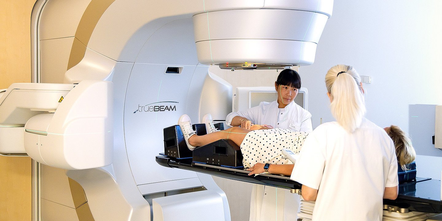
Before treatment begins, specialists carefully determine the individual target volume and develop a radiation treatment plan. The total radiation dose is usually divided into several smaller doses, known as fractions. Depending on the treatment regimen, radiation therapy may be delivered over several days or several weeks, often with up to five sessions per week. A typical session lasts around 20 minutes, although the linear accelerator itself delivers radiation for only a few minutes. During treatment, the patient remains alone in the treatment room, while the medical team continuously monitors them via cameras and can communicate through an intercom system.
“The use of modern radiation therapy techniques, such as intensity-modulated radiation therapy (IMRT) with daily image guidance — known as IGRT — significantly reduces the risk of side effects outside the treatment area.”
— Prof. Robert Krempien, Head of the Department of Radiation Therapy and Radio-Oncology at Helios Hospital Berlin-Buch.
Radiation therapy uses high-energy radiation to damage cancer cells and ultimately destroy them. A linear accelerator precisely directs the radiation toward the tumor area. To minimize side effects, it is important to deliver the required therapeutic dose to the tumor while keeping radiation exposure to the surrounding healthy tissues as low as possible.
Radiation therapy or combined chemoradiotherapy is commonly used for stage II–III rectal cancer before surgery to reduce the tumor volume as much as possible and create more favorable conditions for the operation. Such treatment may also increase the likelihood of preserving the anal sphincter and its function during subsequent surgery.
In selected cases, chemoradiotherapy may be administered after surgery. This may be necessary, for example, if surgery reveals that the disease is more advanced than indicated by the preoperative examinations or if other factors associated with an increased risk of recurrence are identified.
Targeted therapy for colorectal cancer
Targeted therapy is a form of drug treatment that acts on specific molecular mechanisms involved in tumor growth and spread. Unlike conventional chemotherapy, which primarily affects rapidly dividing cells, targeted drugs block specific proteins, receptors, or signaling pathways in cancer cells. In colorectal cancer, this type of treatment is used mainly for advanced or metastatic disease and is often combined with chemotherapy.
To select an appropriate drug, molecular genetic testing of the tumor tissue is performed. Mutations in KRAS, NRAS, and BRAF, as well as other molecular alterations, can play an important role in treatment selection. For example, in tumors without certain RAS mutations, monoclonal antibodies targeting the EGFR receptor may be used. Another approach involves inhibiting the formation of new blood vessels that tumors need for growth, using drugs that target the VEGF signaling pathway. Specific targeted treatment options are also available for selected tumors with a BRAF V600E mutation and certain other rare molecular alterations. This is why molecular profiling of the tumor is particularly important when selecting systemic therapy for metastatic colorectal cancer.
At Helios hospitals, this approach forms part of personalized cancer treatment. For example, at Helios Hospital Emil von Behring, extended tumor profiling may be performed in addition to standard molecular diagnostics when indicated. If additional genetic alterations are identified, specialists assess whether an appropriate precision targeted therapy may be an option. At Helios University Hospital Wuppertal, NGS (Next Generation Sequencing) is among the techniques used for molecular tumor profiling, allowing multiple genes to be analyzed simultaneously to identify potential therapeutic targets.
Radionuclide therapy with lutetium-177
For certain rare tumors of the intestine, targeted radionuclide therapy with lutetium-177 (¹⁷⁷Lu) may be used. This treatment is intended primarily not for typical adenocarcinoma of the colon or rectum, but for well-differentiated neuroendocrine tumors (NETs) whose cells express sufficient levels of somatostatin receptors. Before treatment is considered, the presence of these receptors is confirmed using molecular imaging techniques such as PET/CT.
In peptide receptor radionuclide therapy (PRRT), the radioactive isotope lutetium-177 is linked to a molecule capable of binding to somatostatin receptors on tumor cells. After intravenous administration, the radiopharmaceutical accumulates predominantly in cells expressing the corresponding target and delivers localized radiation that damages the tumor cells. This approach combines molecular targeting with the therapeutic effects of a radionuclide.
Treatment of neuroendocrine tumors with lutetium-177 is also available at Helios Hospital Berlin-Buch. The hospital’s Department of Nuclear Medicine provides diagnostic assessment of somatostatin receptor expression as well as radionuclide therapy for NETs. The decision to use this treatment is made individually based on the characteristics of the tumor and diagnostic findings and may be discussed by a multidisciplinary tumor board.
Important: ¹⁷⁷Lu therapy is not a standard treatment for conventional adenocarcinoma of the colon or rectum. It is intended for selected neuroendocrine tumors with the appropriate molecular characteristics.
Immunotherapy for colorectal cancer
Immunotherapy works differently: it helps the patient’s own immune system recognize and attack cancer cells. In colorectal cancer, this treatment is particularly important for tumors with high microsatellite instability (MSI-H) or deficient DNA mismatch repair (dMMR). These tumors accumulate a large number of genetic alterations, making it easier for the immune system to recognize them as abnormal.
Patients with the appropriate biomarkers may be treated with immune checkpoint inhibitors, particularly drugs targeting the PD-1 pathway and, in certain situations, combinations of immunotherapeutic agents. For this reason, tumor tissue is tested to determine its MSI/MMR status before immunotherapy is considered. For most tumors without these characteristics — namely microsatellite-stable (MSS) and mismatch repair-proficient (pMMR) tumors — standard immunotherapy is significantly less effective. Therefore, a diagnosis of colorectal cancer alone is not sufficient to determine whether immunotherapy is appropriate.
At specialized Helios cancer centers, immunotherapy is part of the range of modern treatments available for gastrointestinal tumors. For example, the Gastrointestinal Tumor Center at Helios Hospital Berlin-Buch offers immunotherapy alongside chemotherapy, antibody therapy, and other systemic and local treatment approaches. New immunotherapy strategies for colorectal cancer are also being investigated there as part of clinical trials.
Follow-up care and rehabilitation
After treatment for colorectal cancer, patients generally undergo follow-up rehabilitation. It usually lasts three weeks and should begin no later than 14 days after the completion of treatment. The starting point for rehabilitation may vary depending on the type of treatment received. After surgery, the end of treatment is generally considered to be the last day of the hospital stay, while for outpatient chemotherapy it is the day of the final infusion.
In addition to follow-up rehabilitation, there is also medical oncological rehabilitation. It can be started within one year after completion of the primary treatment and also usually lasts three weeks. As a rule, both types of rehabilitation are provided on an inpatient basis at a specialized rehabilitation clinic; outpatient or day-patient rehabilitation is possible only in selected cases.
As with the treatment itself, the follow-up program after colorectal cancer depends on whether the tumor was located in the colon or rectum and on the stage at which the disease was diagnosed.
Follow-up for stage I
Follow-up after colorectal cancer treatment can include general oncological surveillance as well as regular colonoscopy.
The extent of follow-up examinations always depends on the tumor stage. For example, after successful removal of stage I colon cancer, extensive routine oncological surveillance may not be necessary, and colonoscopy at recommended intervals may be sufficient.
Follow-up for stages II and III
For stages II and III, follow-up is performed regularly and may include additional examinations alongside colonoscopy. In particular, levels of the tumor marker CEA (carcinoembryonic antigen) may be measured to help detect a possible recurrence at an early stage. Ultrasound examinations may also be performed at specified intervals over a period of five years.
Possible examinations during follow-up
- CEA tumor marker testing: The German S3 guideline recommends measuring CEA levels every six months for at least two years.
- Colonoscopy: Ideally, a complete colonoscopy should be performed before cancer treatment begins. If a colonoscopy was not performed before treatment, the first examination is recommended six months after completion of therapy. If the findings are normal, the examination is subsequently repeated after five years.
- Abdominal ultrasound (abdominal sonography): According to the S3 guideline, ultrasound can be used to detect possible liver metastases and is therefore included in follow-up care after colorectal cancer.
- Chest X-ray: A chest X-ray may be performed as part of follow-up care. In particular, for patients with UICC stage II–III rectal cancer, annual examinations up to the fifth year after completion of treatment may be used for the early detection of lung metastases.
- Computed tomography (CT): According to the cited recommendations, CT is not part of routine regular follow-up examinations. In patients with rectal cancer, it may be performed once approximately three months after completion of treatment to obtain baseline images for comparison with subsequent examinations.
- Sigmoidoscopy: The S3 guideline recommends sigmoidoscopy for patients with rectal cancer who have not received neoadjuvant or adjuvant chemoradiotherapy. In these cases, the examination is performed every six months during the first two years after treatment, for a total of four examinations.
Colorectal cancer treatment at Helios hospitals
At specialized Helios cancer centers, colorectal cancer treatment is based on a multidisciplinary approach. Gastroenterologists, visceral surgeons, oncologists, radiologists, pathologists, radiation oncologists, and nuclear medicine specialists jointly review diagnostic findings at multidisciplinary tumor boards and develop an individualized treatment strategy for each patient. Several centers, including Helios Hospital Berlin-Buch and Helios University Hospital Wuppertal, have colorectal cancer centers certified by the German Cancer Society (DKG).
One of the key areas of expertise is minimally invasive and robot-assisted surgery. For example, at Helios Hospital Erfurt, the da Vinci robotic system is used for bowel surgery. It provides the surgeon with a magnified three-dimensional view of the surgical field and enables highly precise instrument movements, which is particularly valuable during rectal cancer surgery in the confined space of the pelvis. According to the hospital, more than 80% of bowel procedures at the center are performed using robot-assisted techniques.
Personalized oncology is another important focus. At Helios University Hospital Wuppertal, molecular tumor profiling includes techniques such as Next Generation Sequencing (NGS) and liquid biopsy. These methods can identify genetic alterations in the tumor that may serve as targets for targeted therapies. The center also participates in clinical trials of new treatment approaches, giving eligible patients potential access to investigational therapeutic strategies.
Helios Hospital Berlin-Buch offers additional specialized treatment options. Alongside surgery, systemic therapy, and radiation therapy, the hospital has a dedicated Department of Nuclear Medicine. It provides peptide receptor radionuclide therapy (PRRT) with lutetium-177 for selected neuroendocrine tumors of the gastrointestinal tract. This is a highly specialized treatment requiring dedicated radionuclide facilities and expertise and is therefore available only at specialized centers. Before treatment, PET/CT is used to determine whether the tumor expresses somatostatin receptors. If sufficient receptor expression is present, these receptors can then be used to deliver the radioactive agent directly to the tumor cells. However, ¹⁷⁷Lu therapy is intended for selected NETs and is not a standard treatment for conventional adenocarcinoma of the colon or rectum.
Clinical research is also an important part of oncology at Helios. Helios hospitals conduct clinical trials in the field of gastrointestinal tumors, including studies investigating new treatment approaches for colorectal and metastatic colorectal cancer. At the Wuppertal center, participation in clinical research is regarded as an important quality criterion of specialized cancer care and helps facilitate the integration of new therapeutic approaches into clinical practice.
If you are interested in arranging diagnosis or treatment at one of the Helios hospitals, you can submit a request using the contact form on our website or contact us via the email address provided at the top of the website.
Learn more about arranging treatment at Helios hospitals ⟶
Do you need more information about Helios Hospitals or want to schedule your treatment?