Brain tumor treatment: Advances in neuro-oncology
ON THIS PAGE:
- Brain tumor diagnosis
- Types of brain tumors
- Brain tumor treatment options
- Treatment of brain metastases
- Rehabilitation and life after brain tumor treatment
- Brain tumor treatment at Helios hospitals
Brain tumors are abnormal growths that develop in the brain tissue or its surrounding membranes, or spread to the brain from other parts of the body. They can be benign or malignant and vary in their location, growth rate, and impact on the nervous system. Treating these tumors requires exceptional precision, as even a small growth can affect areas of the brain responsible for movement, speech, memory, and other vital functions.
Despite the complexity of these conditions, advances in modern neuro-oncology are opening up new possibilities for treatment. Improvements in neurosurgical techniques, radiation therapy, and molecular diagnostics enable doctors to identify tumor characteristics more accurately and select the most appropriate combination of treatments. This personalized approach can help improve treatment outcomes, achieve long-term disease control, and, in some cases, lead to a complete cure.
The exact causes of most primary brain tumors remain unknown. However, their development is associated with genetic changes in cells that disrupt the normal mechanisms controlling cell division. The risk of developing certain brain tumors may increase with age, although some types are more common in children and young adults. Among the most well-established risk factors are exposure to ionizing radiation, particularly previous radiation therapy to the head, and inherited genetic predispositions associated with rare conditions such as neurofibromatosis, Li-Fraumeni syndrome, and Turcot syndrome. Nevertheless, hereditary cases account for only a small proportion of brain tumors, and most develop in people without any identifiable risk factors. Other commonly discussed factors, including mobile phone use, stress, and lifestyle habits, have not been conclusively established as causes of brain tumors.
How are brain tumors diagnosed?
Symptoms of brain tumors can appear at different stages of the disease and largely depend on the tumor's type, size, growth rate, and location. Slow-growing tumors, such as certain meningiomas and low-grade gliomas, may develop with few or no symptoms for a long time and are sometimes discovered incidentally during examinations for unrelated conditions. In contrast, rapidly growing malignant tumors, such as glioblastoma, can cause noticeable neurological symptoms within just a few weeks. Even a small benign tumor may produce symptoms at an early stage if it is located near critical structures of the brain.
Possible symptoms include persistent or worsening headaches, nausea, visual disturbances, speech difficulties, impaired coordination, muscle weakness, memory problems, and changes in behavior. In some cases, an epileptic seizure may be the first sign of the disease. These symptoms do not necessarily indicate a brain tumor, but their occurrence, particularly if they become progressively worse, requires medical evaluation. Sudden weakness, speech difficulties, or a first-time seizure require urgent medical attention.
In addition to diagnostic examinations prompted by symptoms, preventive monitoring may be recommended in certain cases to detect potential abnormalities before the first signs of disease appear. This primarily applies to people with a hereditary predisposition to brain tumors, particularly those with Li-Fraumeni syndrome, neurofibromatosis, or tuberous sclerosis. An individualized assessment of the need for regular examinations is also important for patients who have previously undergone radiation therapy to the head and those with cancers that have a higher risk of spreading to the brain, such as lung cancer or melanoma. Depending on individual risk factors and the clinical situation, a doctor may recommend periodic MRI scans. However, routine brain tumor screening is generally not necessary for people without symptoms or an increased risk.
Main diagnostic methods
When a brain tumor is suspected, the diagnostic process usually begins with a neurological consultation and an assessment of neurological functions, including reflexes, muscle strength, sensation, coordination, vision, and speech. The following methods are used to establish or clarify the diagnosis:
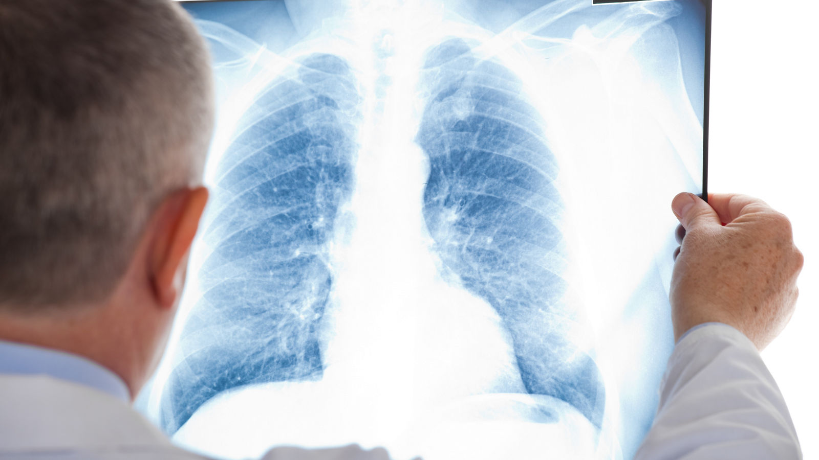
- Magnetic resonance imaging (MRI) is the primary imaging method for detecting and evaluating brain tumors. It helps determine the tumor's location, size, structure, and relationship to surrounding tissues. Contrast-enhanced MRI provides more detailed information about the tumor's characteristics and boundaries.
- Computed tomography (CT) is mainly used when rapid imaging is required, such as in emergency situations, and to detect hemorrhage, calcifications, and changes in the bony structures of the skull.
- Advanced MRI techniques, including functional MRI, diffusion tensor tractography, perfusion MRI, and MR spectroscopy, may be used when indicated to further characterize the tumor and assist in planning surgical treatment.
Additional diagnostic tests for suspected malignant brain tumors
If imaging findings suggest that a tumor may be malignant, examination of tumor tissue is often necessary to establish an accurate diagnosis and select the most appropriate treatment. Tissue samples can be obtained during surgical tumor removal or through a stereotactic biopsy, a minimally invasive procedure in which a small tissue sample is collected using image-guided navigation.
The collected tissue undergoes histopathological examination and, when indicated, molecular genetic testing. Modern diagnostic assessment of gliomas may include testing for IDH1/IDH2 mutations, MGMT promoter methylation status, and 1p/19q codeletion. These molecular markers help determine the precise tumor type, assess prognosis, and identify which treatments are most likely to be effective.
In certain clinical situations, positron emission tomography (PET), including PET with amino acid radiotracers, may also be performed. This technique provides additional information about the tumor's metabolic activity, helps identify the most appropriate area for biopsy, or assists in distinguishing active tumor tissue from treatment-related changes.
The final diagnosis is based on a combination of clinical findings, imaging results, and, when necessary and feasible, examination of tumor tissue. This comprehensive diagnostic approach enables specialists to develop the most appropriate treatment strategy based on the individual characteristics of the disease.
Types of brain tumors
Brain tumors are classified according to their origin, cell type, molecular characteristics, and grade. They are broadly divided into primary tumors, which develop within the brain or its surrounding structures, and secondary (metastatic) tumors, which arise when cancer cells spread to the brain from other parts of the body. Primary brain tumors can be benign or malignant. According to the World Health Organization (WHO) classification, many central nervous system tumors are assigned a CNS WHO grade ranging from 1 to 4, reflecting their biological behavior and helping doctors determine the most appropriate treatment strategy.
Gliomas
Gliomas are a broad group of primary central nervous system tumors that include astrocytomas, oligodendrogliomas, glioblastomas, and other types of tumors. They differ in their growth rate, molecular characteristics, and grade.
Astrocytomas can vary considerably in their degree of malignancy, ranging from relatively slow-growing forms to aggressive tumors requiring comprehensive treatment. Oligodendrogliomas typically grow relatively slowly and are classified as CNS WHO grade 2 or 3 tumors. Their defining molecular features include an IDH mutation and 1p/19q codeletion.
Glioblastoma is one of the most aggressive primary brain tumors in adults and is classified as CNS WHO grade 4. It is characterized by rapid growth and the ability to infiltrate surrounding brain tissue. As a result, treatment usually involves a combination of surgery, radiation therapy, and drug therapy.
Meningiomas
Meningiomas develop from cells of the meninges, the protective membranes surrounding the brain and spinal cord, and are among the most common primary central nervous system tumors in adults. Most are low-grade tumors (CNS WHO grade 1), grow slowly, and may remain asymptomatic for a long time.
However, more aggressive forms also occur. Grade 2 and 3 meningiomas are associated with a higher risk of recurrence. Depending on the tumor's location, size, and growth characteristics, treatment may involve active surveillance, surgical removal, or radiation therapy.
Pituitary tumors
Pituitary tumors develop in the pituitary gland, which regulates various functions of the endocrine system. The most common are pituitary adenomas, also referred to as pituitary neuroendocrine tumors (PitNETs) in modern classification systems. Most have a benign clinical course, although some may exhibit aggressive growth.
These tumors can cause hormonal imbalances, headaches, and visual disturbances due to pressure on nearby structures. Treatment depends on the tumor's size, hormonal activity, and clinical manifestations. Depending on the specific tumor type, treatment options may include medication, surgery, or radiation therapy.
Ependymomas
Ependymomas are central nervous system tumors associated with ependymal cells, which line the ventricles of the brain and the central canal of the spinal cord. They occur in both children and adults, although their location and molecular characteristics may vary with age.
Some ependymomas grow relatively slowly, while others follow a more aggressive course and carry a higher risk of recurrence. The primary treatment is usually maximal safe surgical removal of the tumor, which may be followed by radiation therapy when indicated.
Brain metastases
Brain metastases are secondary malignant tumors that develop when cancer cells spread to the brain from other parts of the body. They most commonly originate from lung cancer, breast cancer, melanoma, kidney cancer, and certain other malignancies.
Metastatic lesions may be single or multiple, and their clinical manifestations depend on the size, location, and number of lesions. Unlike primary brain tumors, treatment decisions for brain metastases depend not only on the characteristics of the brain lesions but also on the type of primary cancer, the extent of the disease, and the patient's overall condition. Treatment may include surgical removal, stereotactic radiation therapy, and systemic anticancer therapies.
Brain tumor treatment options
Brain tumor treatment is tailored to each patient, taking into account the tumor's type, location, size, grade, and molecular characteristics, as well as the patient's overall health. Modern neuro-oncology offers a range of treatment options, including surgical removal, radiation therapy, chemotherapy, targeted therapies, and other specialized approaches. Depending on the diagnosis, these methods may be used individually or in combination. The primary goal is not only to achieve the best possible disease control but also to preserve neurological function and the patient's quality of life.
Surgical removal of brain tumors
Surgery plays a key role in treating many primary brain tumors, including gliomas, meningiomas, and certain pituitary tumors, as well as selected cases of brain metastases. Depending on the clinical situation, surgery may aim to remove the tumor completely, reduce its size, or relieve pressure on surrounding brain structures. Tissue obtained during the procedure also helps establish a precise diagnosis and enables molecular testing to guide further treatment.
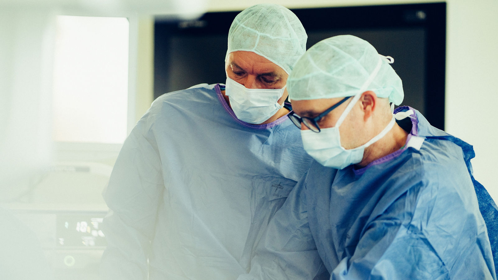
The guiding principle of modern neuro-oncological surgery is maximal safe resection. This means that the neurosurgeon aims to remove as much tumor tissue as possible while minimizing the risk of damage to brain regions responsible for speech, movement, vision, and other essential functions. To achieve this, surgeons use advanced imaging, navigation, and intraoperative monitoring technologies.
“When operating on the brain, there is no room for error,” — Prof. Dr. med. Barbara Carl, Neurosurgeon, Helios Dr. Horst Schmidt Hospital Wiesbaden.
Microsurgery, neuronavigation, and augmented reality
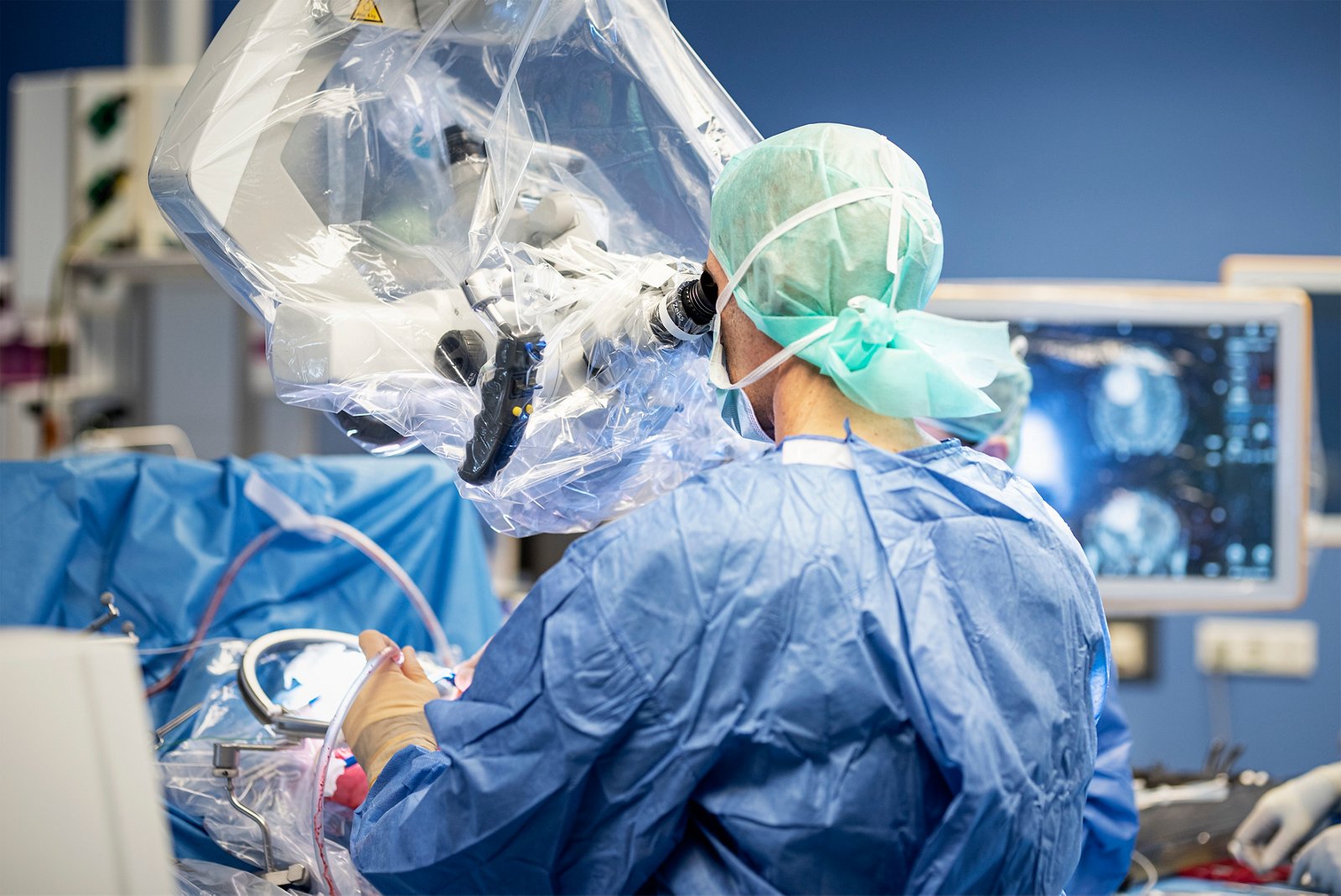
Microsurgical tumor removal is performed using an operating microscope, which provides high magnification and detailed visualization of blood vessels, nerves, and surrounding tissues. This allows neurosurgeons to operate with a high degree of precision, even in complex anatomical areas where tumors are located close to critical brain structures.
Neuronavigation provides additional precision by matching the position of surgical instruments with MRI or CT images obtained before surgery. This technology helps surgeons plan the optimal approach to the tumor and navigate the brain's anatomy during the procedure. A further development of this approach is augmented reality (AR), which allows three-dimensional representations of anatomical structures to be overlaid onto the surgical field. This helps surgeons better understand the tumor's position in relation to blood vessels, neural pathways, and functionally important brain regions.
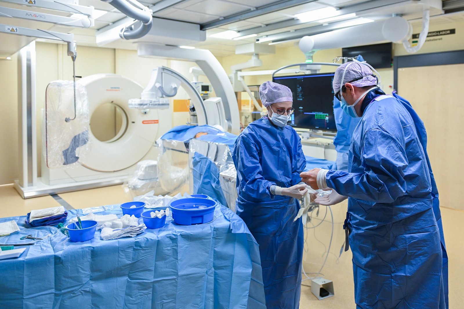
Advanced microsurgical and neuronavigation techniques are used at Helios Dr. Horst Schmidt Hospital (HSK) Wiesbaden and Helios Hospital Berlin-Buch. In Wiesbaden, specialists use augmented reality during brain surgery, while neurosurgical procedures at Berlin-Buch are performed with operating microscopes, navigation systems, and advanced imaging technologies.
Fluorescence-guided brain tumor surgery
One of the main challenges in removing certain malignant brain tumors, particularly high-grade gliomas, is the lack of a clearly defined boundary between tumor tissue and healthy brain tissue. To help identify areas that need to be removed more precisely, surgeons may use fluorescence-guided surgery with 5-aminolevulinic acid (5-ALA).
Before surgery, the patient takes a special medication that promotes the accumulation of fluorescent compounds in certain tumor cells. During the procedure, the surgeon uses a microscope equipped with a special illumination mode to detect the characteristic fluorescence of tumor tissue. This makes it easier to identify tumor areas that may be difficult to distinguish from surrounding tissue under normal lighting and supports more complete tumor removal within the limits of safe resection.
Fluorescence-guided surgery using 5-ALA is among the neuro-oncological technologies available at Helios Hospital Krefeld and Helios Hospital Erfurt. At both centers, the technique is used when clinically indicated to provide more precise visualization of tumor tissue during surgery.
Awake brain surgery and intraoperative neuromonitoring
When a tumor is located near brain regions responsible for speech, movement, or other essential functions, surgery requires particularly careful planning. In such cases, awake brain surgery may be considered, allowing surgeons to assess certain neurological functions directly during tumor removal.
During specific stages of the procedure, the patient remains conscious and performs simple tasks, such as naming objects, speaking, or answering questions. Pain is controlled through anesthesia, and this approach is used only when clinically appropriate. The technique helps surgeons identify functionally important brain regions and reduce the risk of postoperative speech difficulties and other neurological impairments.
Another important technology is intraoperative neuromonitoring (IONM). By recording and assessing electrical signals, specialists monitor motor, sensory, and certain neural pathways during surgery. Unlike awake brain surgery, neuromonitoring can also be performed under general anesthesia. These techniques often complement each other, helping surgeons remove tumors in functionally important brain regions as safely as possible.
Awake brain surgery and intraoperative neuromonitoring are available in the neurosurgical departments of Helios Hospital Schwerin and Helios Hospital Bad Saarow. Both centers use function-preserving surgical approaches designed to maximize tumor removal while protecting neurological function.
Intraoperative imaging: MRI and ultrasound
Even with careful preoperative planning, the position and shape of brain tissues may change during surgery, reducing the accuracy of navigation systems based solely on previously acquired images. To provide additional guidance, neurosurgeons may use intraoperative imaging to assess the location and extent of residual tumor tissue during the procedure.
Intraoperative MRI (iMRI) provides updated images of the brain during surgery. It can help detect remaining tumor tissue and determine whether further removal is possible without compromising patient safety. Intraoperative ultrasound also provides real-time imaging, helping surgeons assess tumor boundaries and monitor the progress of resection. The choice of technology depends on the tumor type, the surgical procedure, and the equipment available at the neurosurgical center.
Intraoperative MRI is used at Helios Hospital Erfurt, including in the context of neuro-oncological research involving glioblastoma. At Helios Hospital Krefeld, intraoperative ultrasound is also used to visualize tumor tissue in selected clinical situations.
Minimally invasive and endoscopic skull base surgery
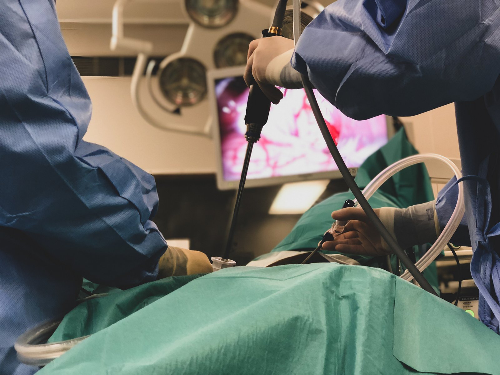
Certain tumors, particularly those affecting the pituitary gland and skull base, can be removed using endoscopic or other minimally invasive surgical approaches. Unlike conventional open surgery, endoscopic procedures use a thin instrument equipped with a camera and light source, allowing surgeons to visualize difficult-to-reach anatomical areas through relatively small surgical openings.
For certain pituitary tumors, an endoscopic endonasal approach may be used, allowing the surgeon to reach the tumor through the nasal passages and sphenoid sinus. This technique can avoid the need for a large craniotomy (surgical removal of a section of the skull), although it is not suitable for every tumor. Its feasibility depends on the tumor's size, location, and extent, as well as its proximity to major blood vessels and the optic nerves.
Endoscopic procedures for pituitary and skull base tumors are part of the neurosurgical services offered at Helios Hospital Erfurt, Helios Dr. Horst Schmidt Hospital (HSK) Wiesbaden, and Helios Hospital Krefeld. In Erfurt, surgeons use three-dimensional endoscopy for skull base procedures, while in Krefeld, neuroendoscopic techniques are also employed in the treatment of certain pituitary tumors.
Radiation therapy for brain tumors
Radiation therapy is one of the main treatment options for brain tumors. It uses high-energy radiation to damage tumor cells and inhibit their growth. It may be used as a standalone treatment, for example when surgical removal is not possible or carries significant risks, or in combination with surgery and drug therapy. Following surgery, radiation therapy is often recommended to target remaining tumor cells and reduce the risk of recurrence.
Radiation therapy is used to treat various types of brain tumors, including glioblastomas, certain other gliomas, meningiomas, and brain metastases. Modern technologies allow specialists to calculate radiation doses precisely and target specific areas while minimizing exposure to surrounding healthy tissues. The choice of technique, treatment duration, and radiation schedule depends on the diagnosis, tumor size and location, previous treatments, and the patient's individual circumstances.
Precision radiation therapy: IMRT and VMAT
One of the most widely used modern approaches is intensity-modulated radiation therapy (IMRT). This technique adjusts the intensity of radiation within individual beams, allowing the dose distribution to conform to the complex shape of a tumor while reducing exposure to surrounding brain structures.
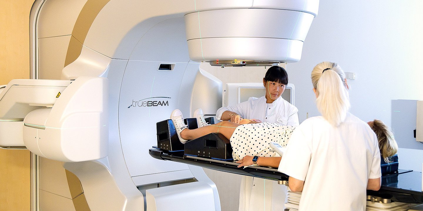
A further development of this approach is volumetric modulated arc therapy (VMAT). During treatment, a linear accelerator rotates around the patient while continuously adjusting the radiation intensity and beam shape. This enables precise dose distribution and, in some cases, shorter treatment sessions. Image-guided radiation therapy (IGRT) may also be used to improve accuracy by checking the patient's positioning with imaging immediately before treatment.
IMRT and VMAT are particularly valuable when treating tumors located near the optic nerves, brainstem, and other sensitive structures. For malignant gliomas, radiation therapy is usually delivered over a series of sessions, allowing the prescribed dose to be distributed over time.
Advanced IMRT and VMAT technologies are used at Helios Park Hospital Leipzig, where radiation therapy is also part of the treatment options for brain tumors. At Helios Hospital Bad Saarow, specialists use IMRT and image-guided techniques to precisely target tumor tissue while taking nearby organs and anatomical structures into account.
Stereotactic radiotherapy
Stereotactic radiotherapy (SRT) is a highly precise radiation treatment technique that directs radiation to a small, carefully defined target using specialized treatment planning and patient positioning systems. Its high spatial accuracy allows a substantial radiation dose to be delivered to the tumor while limiting exposure to surrounding tissues.
Stereotactic radiation therapy may be used for selected meningiomas, vestibular schwannomas, brain metastases, and certain other tumors. When treatment is delivered over several sessions, it is known as fractionated stereotactic radiotherapy. This approach may be preferable for larger tumors or those located close to sensitive structures, as dividing the radiation dose into multiple sessions can help reduce the risk of damage to healthy tissues.
At Helios Hospital Bad Saarow, high-precision stereotactic radiotherapy is used for selected benign and malignant brain tumors, including metastases. Specialists at Helios Hospital Schwerin also provide intracranial stereotactic radiotherapy, including treatment for meningiomas, vestibular schwannomas, and metastatic brain lesions.
Stereotactic radiosurgery: Gamma Knife and CyberKnife
Stereotactic radiosurgery (SRS) is a specialized form of high-precision radiation therapy in which a high dose of radiation is delivered to a tumor in a single session. Despite its name, radiosurgery does not involve a surgical incision. Its purpose is to achieve local tumor control without open surgery.
Different systems are used to perform radiosurgery. Gamma Knife directs multiple narrow beams of gamma radiation toward a precisely defined target, where the beams converge. CyberKnife is a robotic radiation therapy system consisting of a compact linear accelerator mounted on a robotic arm. This allows radiation to be delivered from multiple angles with high precision. Depending on the clinical situation, treatment with these systems may be delivered in one or several sessions.
Radiosurgery may be considered for small brain metastases, certain meningiomas, vestibular schwannomas, and other well-defined lesions. However, it is not a universal alternative to surgical removal. The choice of treatment depends on the tumor type, size, location, and therapeutic goals.
At Helios Hospital Krefeld, the neuro-oncology team collaborates with a local Gamma Knife center, considering radiosurgery as a treatment option when clinically appropriate. At Helios Hospital Erfurt, high-precision radiosurgery using CyberKnife is available in collaboration with Cyberknife Centrum Mitteldeutschland.
Whole-brain radiation therapy
In certain cases, primarily when multiple brain metastases are present, whole-brain radiation therapy (WBRT) may be recommended. Unlike stereotactic techniques that target individual lesions, this approach delivers radiation to the brain as a whole, allowing treatment of both visible metastases and potentially microscopic tumor deposits.
However, WBRT may be associated with more pronounced long-term side effects, including memory problems and other cognitive impairments. When choosing between whole-brain radiation and localized treatment methods, specialists therefore consider the number and total volume of metastases, the characteristics of the primary cancer, the patient's prognosis, and overall health. When WBRT is necessary, hippocampal avoidance techniques and medication to help preserve cognitive function may be used in selected patients.
At Helios Hospital Berlin-Buch, brain tumors and metastases are treated as part of comprehensive radiation oncology care. Decisions regarding the extent of radiation treatment and its combination with other therapies are made individually, taking the specific characteristics of each patient's disease into account.
Chemotherapy for brain tumors
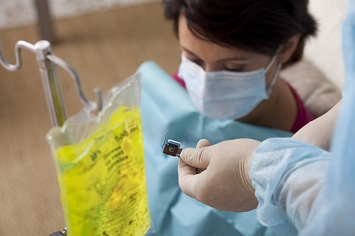
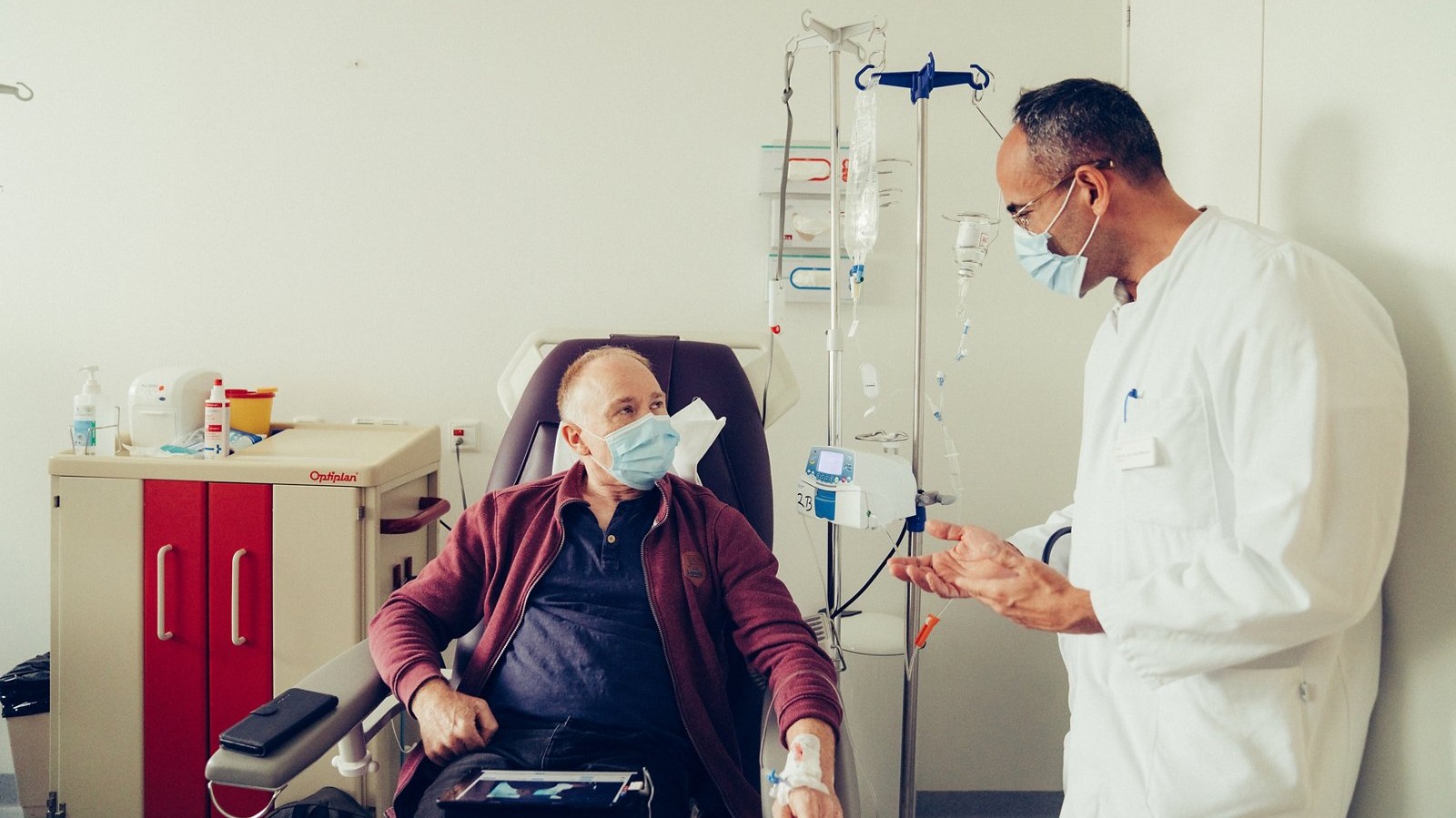
Chemotherapy is used to inhibit the growth and division of tumor cells and plays an important role in treating certain malignant brain tumors, particularly gliomas. Depending on the diagnosis, it may be administered after surgery, concurrently with radiation therapy, or when the disease progresses. The choice of medication depends not only on the tumor type but also on its molecular characteristics, previous treatments, and the patient's overall health.
One of the main chemotherapy drugs used in neuro-oncology is temozolomide. It is widely used to treat glioblastoma, usually in combination with radiation therapy, followed by additional cycles of chemotherapy. Treatment effectiveness may depend on the tumor's molecular characteristics, particularly the methylation status of the MGMT gene promoter, which helps predict the likelihood of response to temozolomide.
For certain types of gliomas, doctors also use the PCV chemotherapy regimen, which combines procarbazine, lomustine, and vincristine. This regimen is particularly important for certain oligodendrogliomas and other gliomas for which clinical studies have demonstrated a treatment benefit. Lomustine may also be used on its own, particularly in selected cases of recurrent malignant glioma.
One of the challenges of treating brain tumors with medication is the blood-brain barrier, a natural protective mechanism that limits the passage of many substances from the bloodstream into brain tissue. As a result, not all anticancer drugs can reach sufficient concentrations within the tumor, making it essential to select chemotherapy based on the characteristics of each patient's disease.
Throughout treatment, doctors regularly assess its effectiveness using MRI scans and monitor potential side effects, including low blood cell counts, nausea, and fatigue. The treatment regimen and duration may be adjusted as needed to achieve the best possible balance between anticancer effectiveness, treatment tolerability, and the patient's quality of life.
Targeted therapy
Targeted therapy is a modern approach to cancer treatment that focuses on specific molecular mechanisms involved in the growth of tumor cells. Unlike traditional chemotherapy, which primarily affects rapidly dividing cells, targeted therapies act on specific molecular targets. To determine whether a patient may benefit from this treatment, specialists perform molecular genetic testing of tumor tissue to identify relevant mutations and other biological characteristics of the tumor.
One of the major advances in recent years has been the development of drugs targeting mutations in the IDH1 and IDH2 genes, which are found in certain types of gliomas. In particular, vorasidenib blocks the activity of mutated IDH enzymes and helps slow tumor growth. In the European Union, it is approved for certain CNS WHO grade 2 astrocytomas and oligodendrogliomas with these mutations in patients who have undergone surgery and do not yet require immediate radiation therapy or chemotherapy. For these patients, targeted treatment may help delay disease progression and the need for other anticancer treatments.
Another example of a personalized treatment approach involves tumors carrying the BRAF V600E mutation. This genetic alteration is found in certain gliomas, including some childhood brain tumors. In such cases, drugs that block signaling pathways involved in tumor cell growth may be used, such as the combination of dabrafenib and trametinib. In the EU, this combination is approved for certain BRAF V600E-mutated gliomas in children. Whether it can be used depends on the patient's age, tumor type, previous treatments, and approved indications.
Despite advances in targeted therapy, these medications are not suitable for all brain tumors and do not automatically replace surgery, radiation therapy, or chemotherapy. Treatment decisions are made individually, taking into account the tumor's molecular profile, the results of previous treatments, and the available clinical evidence.
At Helios Hospital Erfurt, a personalized approach to brain tumor treatment combines advanced molecular genetic testing, neuropathological diagnostics, and multidisciplinary discussion of each patient's case. The center also participates in the VIOLETA study, which evaluates the effectiveness, safety, and quality of life of patients with IDH-mutant grade 2 gliomas receiving vorasidenib in routine clinical practice.
Tumor Treating Fields (TTFields)
Tumor Treating Fields (TTFields) is a modern, non-invasive treatment primarily used for patients with glioblastoma. Unlike radiation therapy, which uses ionizing radiation, TTFields delivers low-intensity, alternating electric fields at intermediate frequencies to tumor cells. These fields interfere with cell division and may help slow tumor growth.
Treatment is delivered using a portable system called Optune. Special electrode arrays are placed on the patient's shaved scalp and connected to a small device that generates electric fields. The therapy is administered continuously for most of the day, including at home, allowing patients to maintain many of their usual daily activities. For optimal effectiveness, patients are generally advised to use the device for at least 18 hours per day.
For newly diagnosed glioblastoma, TTFields may be used after completion of radiation therapy alongside maintenance chemotherapy with temozolomide. Clinical studies have shown that adding TTFields to standard treatment can extend overall survival and the time before disease progression in selected patients. However, the treatment is not suitable for everyone. Decisions take into account the diagnosis, the patient's overall health, previous treatments, and their ability to use the device consistently.
Because TTFields does not cause the same systemic effects as chemotherapy, its side effects are generally associated with the electrode arrays. The most common include scalp irritation, redness, and itching. An important consideration is the need for prolonged daily use, making patient education and ongoing support from the medical team essential.
Specialists at Helios Hospital Erfurt have experience studying TTFields through the TIGER study, which investigated the use of this technology in patients with newly diagnosed glioblastoma in routine clinical practice. Helios Hospital Schwerin also participates in research related to this treatment, including the Cocoon project, which focuses on improving patient adherence to TTFields therapy. At Helios Hospital Krefeld, neuro-oncology specialists consider TTFields as an additional treatment option for glioblastoma. Whether this technology is appropriate for an individual patient is determined as part of a comprehensive, personalized treatment plan.
Immunotherapy and emerging treatments
Immunotherapy is one of the most promising areas of modern neuro-oncology, aiming to harness the patient's own immune system to fight tumor cells. Unlike chemotherapy, which directly affects cell division, immunotherapeutic approaches help the immune system recognize and attack tumors. However, immunotherapy remains particularly challenging for primary brain tumors, especially glioblastoma. This is partly due to the unique immune environment surrounding these tumors and their ability to suppress the body's antitumor immune response.
One of the most extensively studied approaches involves immune checkpoint inhibitors — drugs that block signals preventing immune cells from attacking tumors. These medications have already proven effective against several types of cancer. However, large clinical trials have not yet demonstrated a clear benefit from their routine use in glioblastoma. As a result, they are not part of standard treatment for most primary brain tumors and are mainly considered within clinical trials or in selected cases involving specific molecular characteristics.
Another promising area is the development of cancer vaccines, designed to train the immune system to recognize specific antigens on tumor cells. In particular, researchers are investigating dendritic cell vaccines, which use the patient's own immune cells prepared to recognize tumor antigens. Personalized vaccines tailored to the molecular characteristics of an individual tumor are also being developed. Despite encouraging findings from some studies, these approaches have not yet become standard treatments for glioblastoma, and further research is needed to confirm their effectiveness.
Research is also underway in cell-based immunotherapy, including CAR-T cell therapy. This approach involves genetically modifying a patient's own T lymphocytes so they can recognize specific molecules on the surface of tumor cells. CAR-T cell therapy is already used to treat certain blood cancers, but its application to brain tumors remains experimental. Key challenges include identifying suitable molecular targets, overcoming the tumor's protective mechanisms, and ensuring treatment safety.
At Helios hospitals, modern immunotherapy approaches are used to treat various types of cancer, while specialists are also involved in research into new treatments for primary brain tumors. In particular, Helios Hospital Krefeld is among the centers participating in the GlioVax clinical trial, which investigates the use of a dendritic cell vaccine in combination with standard radiation therapy and chemotherapy for patients with newly diagnosed glioblastoma. The study aims to evaluate the safety and effectiveness of this approach, including its potential impact on patient survival.
Although many immunotherapeutic approaches for primary brain tumors are still being investigated in clinical trials, their development offers new opportunities to improve treatment. Research into the molecular characteristics of tumors and their interactions with the immune system is helping scientists develop more precise therapeutic strategies and identify which patients may benefit most from them.
Treatment of brain metastases
Brain metastases are secondary malignant tumors that develop when cancer cells spread to the brain from other parts of the body. They most commonly occur in patients with lung cancer, breast cancer, melanoma, and certain other cancers. Unlike primary brain tumors, treating brain metastases requires consideration not only of the condition of the brain but also of the characteristics of the primary cancer, the extent of disease throughout the body, and the effectiveness of previous treatments.
The choice of treatment depends on the number, size, and location of the metastases, the presence of neurological symptoms, the molecular characteristics of the primary tumor, and the patient's overall health. Decisions are made by a multidisciplinary team of specialists, who determine the most appropriate combination of local treatments targeting brain metastases and systemic anticancer therapy.
Surgical removal may be recommended for selected large or symptomatic metastases, particularly when they can be safely accessed. Surgery can relieve pressure on surrounding brain structures, improve neurological symptoms, and provide tissue for a more precise diagnosis. Following surgery, stereotactic radiation therapy is often delivered to the surgical cavity to reduce the risk of local recurrence.
Stereotactic radiosurgery and radiotherapy are widely used to treat a limited number of brain metastases. In selected cases, they may also be suitable for multiple lesions, provided their total volume and location allow treatment to be delivered safely. These techniques deliver highly precise radiation to tumor sites while minimizing exposure to surrounding healthy tissue. For more extensive brain involvement, whole-brain radiation therapy may be considered. However, because of the risk of cognitive impairment, the decision takes into account the expected benefits, the patient's prognosis, and available alternatives.
Systemic drug therapy also plays an important role in treating the underlying cancer. Depending on the type and molecular characteristics of the primary tumor, treatment may include targeted therapy, immunotherapy, or chemotherapy. Some modern anticancer drugs can also be effective against brain metastases. For example, certain targeted therapies are used for lung cancers with EGFR mutations or ALK rearrangements, as well as HER2-positive breast cancer. Immunotherapy may benefit selected patients with melanoma and certain types of lung cancer. When choosing treatment, specialists consider the drugs' demonstrated activity in the central nervous system and the need to control cancer elsewhere in the body.
If brain swelling causes symptoms, corticosteroids may be prescribed temporarily to reduce swelling and pressure on surrounding tissues. Antiepileptic medications are used when seizures occur but are generally not prescribed preventively to patients who have never experienced a seizure. Supportive care helps relieve symptoms and maintain quality of life during anticancer treatment.
After the initial course of treatment, patients require regular follow-up with brain MRI scans and assessment of the underlying cancer. If new lesions appear or previously treated metastases begin to grow again, additional local or systemic treatments may be considered. Modern multidisciplinary care allows many patients to achieve long-term control of brain metastases, reduce neurological symptoms, and maintain their ability to carry out everyday activities.
Rehabilitation and life after brain tumor treatment
Recovery after brain tumor treatment depends on the tumor's location, size, and characteristics, as well as the treatments performed. Following surgery, some patients return to their usual activities relatively quickly, while others require a longer period of rehabilitation. Possible effects include muscle weakness, impaired coordination, speech difficulties, problems with memory and concentration, and increased fatigue. Some neurological symptoms may gradually improve as the patient recovers from surgery.
Neurorehabilitation focuses on restoring lost functions, improving independence, and enhancing quality of life. Depending on the patient's needs, a rehabilitation program may include physiotherapy to improve movement and balance, occupational therapy to support everyday activities, speech and language therapy for speech or swallowing difficulties, and neuropsychological support for problems with memory, attention, or emotional well-being. Whenever possible, rehabilitation begins early in the postoperative period and is adjusted as the patient's condition improves.
Returning to work, physical exercise, and other everyday activities is usually a gradual process. The timeline depends on the patient's overall condition, any remaining neurological symptoms, and the need for further treatment. Driving and activities involving an increased risk of injury require particular attention, as temporary restrictions may apply after seizures or certain neurosurgical procedures. Adequate sleep, moderate physical activity, and psychological support for patients and their families also play an important role in recovery.
After brain tumor removal, regular medical follow-up is essential, even if surgery was successful and the patient feels significantly better. Follow-up MRI scans help assess postoperative changes and detect any residual tumor growth or recurrence at an early stage. The frequency of examinations depends on the tumor type, treatment results, and individual risk of recurrence. For malignant tumors, follow-up also includes monitoring the effectiveness of additional radiation therapy or drug treatment.
Modern brain tumor care goes beyond removing the tumor or controlling its growth. An equally important goal is to help patients regain as much independence as possible, preserve their cognitive and physical abilities, and return to a fulfilling everyday life.
Brain tumor treatment at Helios hospitals
Helios hospitals in Germany offer a comprehensive approach to brain tumor diagnosis and treatment, bringing together the expertise of neurosurgeons, neuro-oncologists, radiation oncologists, and other specialists. Depending on the diagnosis and individual characteristics of the disease, treatment may involve advanced neuroimaging, microsurgical and minimally invasive procedures, high-precision radiation therapy, and various forms of drug therapy. Multidisciplinary collaboration enables specialists to develop personalized treatment plans with particular attention to preserving neurological function and patients' quality of life.
“To provide our patients with comprehensive, high-quality care, neurosurgeons, neurologists, radiation oncologists, oncologists, neuroradiologists, nuclear medicine specialists, and other experts work closely together at our center,” — Prof. Dr. med. Rüdiger Gerlach, Neurosurgeon, Helios Hospital Erfurt.
For international patients, Helios International provides support throughout the process of arranging treatment in Germany, from the initial review of medical records and selection of a suitable hospital to coordinating the diagnostic or treatment program.
If you are interested in arranging diagnosis or treatment at one of the Helios hospitals, you can submit a request using the contact form on our website or contact us via the email address provided at the top of the website.
Learn more about arranging treatment at Helios hospitals ⟶
Do you need more information about Helios Hospitals or want to schedule your treatment?