
Stomach cancer: diagnosis and modern treatment methods
- Causes and risk factors for stomach cancer
- Stomach cancer diagnosis
- Stomach cancer treatment methods
- Life after stomach cancer treatment
- Stomach cancer treatment at Helios Hospitals
Stomach cancer is among the five most common cancers worldwide. According to the International Agency for Research on Cancer (IARC), approximately 980,000 new cases were reported in 2024. Despite advances in modern treatment methods, stomach cancer remains one of the leading causes of cancer-related deaths.
The most common type of stomach cancer is adenocarcinoma, accounting for approximately 90–95% of cases. Based on the characteristics of the tumor cells, gastric adenocarcinomas are classified into intestinal and diffuse types. The diffuse type generally tends to be more aggressive and more likely to spread rapidly. Less common types include lymphomas, gastrointestinal stromal tumors (GISTs), and neuroendocrine tumors. The choice of treatment depends on the tumor type, its stage, molecular characteristics, and the patient's overall health.
One of the main challenges of stomach cancer is that it often develops without noticeable symptoms in its early stages or causes only mild discomfort that can easily be mistaken for common digestive problems. However, early detection significantly improves the chances of successful treatment and, in some cases, allows the tumor to be removed endoscopically without major surgery. This is why it is important not to ignore persistent or recurring stomach symptoms, seek timely advice from a gastroenterologist, and undergo appropriate examinations, especially if risk factors are present.
Causes and risk factors for stomach cancer
Stomach cancer develops as a result of genetic changes in the cells of the stomach lining, leading to uncontrolled cell growth and the formation of a malignant tumor. In most cases, it is impossible to identify a single specific cause of the disease. However, several factors can significantly increase the risk of developing stomach cancer.
The main risk factors include:
- Helicobacter pylori infection. One of the most significant risk factors for stomach cancer. Long-term infection can cause chronic inflammation of the stomach lining, leading to atrophy and precancerous changes. However, most people infected with H. pylori do not develop stomach cancer.
- Chronic stomach conditions. Atrophic gastritis, intestinal metaplasia, and dysplasia of the stomach lining increase the risk of malignant transformation. Pernicious anemia, which is associated with autoimmune gastritis, may also increase the risk.
- Hereditary predisposition. The risk of stomach cancer is higher in people whose close relatives have had the disease. In rare cases, tumor development is associated with inherited genetic syndromes, including hereditary diffuse gastric cancer linked to mutations in the CDH1 gene.
- Dietary habits. Frequent consumption of heavily salted, smoked, and salt-preserved foods is associated with an increased risk of stomach cancer. Low consumption of fresh fruits and vegetables may also play a role.
- Smoking. Tobacco smoke contains carcinogenic substances that increase the risk of developing malignant tumors, including stomach cancer. The risk increases with the intensity and duration of smoking.
- Age and sex. The likelihood of developing stomach cancer increases with age. The disease is more commonly diagnosed in people over 60, and men are approximately twice as likely to develop it as women.
- Overweight and obesity. These factors are particularly associated with an increased risk of cancer of the gastric cardia, the area where the stomach connects to the esophagus.
- Previous stomach surgery. After partial removal of the stomach for benign conditions, the risk of developing a malignant tumor in the remaining portion of the organ may increase many years later.
- Epstein–Barr virus (EBV) infection. A small proportion of stomach cancer cases are associated with this virus. However, having an EBV infection does not necessarily mean that a person will develop a malignant tumor.
Having one or more risk factors does not mean that stomach cancer will inevitably develop. Nevertheless, people with a hereditary predisposition, precancerous changes in the stomach lining, or persistent stomach symptoms should discuss the need for appropriate examinations and individualized monitoring with their doctor.
Stomach cancer diagnosis
Early diagnosis of stomach cancer plays a crucial role in successful treatment. In its early stages, the tumor often causes no noticeable symptoms, making it difficult to detect without specific examinations. The primary diagnostic method is gastroscopy, which allows doctors to examine the stomach lining and, if necessary, collect tissue samples for further analysis.
Preventive screening: who should have their stomach checked and how often?
Regular gastroscopy is not a mandatory examination for everyone. The need for preventive screening depends on age, family history, chronic stomach conditions, and the prevalence of stomach cancer in the country of residence. For example, some East Asian countries, where the disease is particularly common, have national screening programs. People without symptoms or increased risk factors generally do not need annual gastroscopy.
The following groups should pay particular attention to their stomach health:
- People with a family history of stomach cancer. If a parent, sibling, or child has been diagnosed with the disease, it is advisable to discuss the need for gastroscopy with a gastroenterologist. Genetic counseling may be necessary if a hereditary cancer syndrome is suspected.
- Patients with atrophic gastritis or intestinal metaplasia. These changes in the stomach lining may increase the risk of developing a malignant tumor. In cases of extensive or severe changes, follow-up gastroscopy is often recommended approximately every three years. However, the exact interval depends on biopsy results and additional risk factors.
- People with hereditary genetic syndromes. Certain inherited conditions, such as Lynch syndrome or hereditary diffuse gastric cancer, may require a specialized surveillance program. The frequency of examinations is determined individually at a specialized medical center.
- Patients who have undergone stomach surgery. After partial removal of the stomach, particularly for benign conditions, the risk of developing a malignant tumor in the remaining portion may increase many years later. The need for regular gastroscopy depends on the type of surgery, the time elapsed since the procedure, and individual risk factors.
- People with Helicobacter pylori infection. If the bacterium is detected, patients should discuss eradication therapy with their doctor to eliminate the infection. After treatment, testing is needed to confirm that the infection has been successfully eradicated. However, H. pylori infection alone does not necessarily mean that regular gastroscopy is required.
If warning symptoms appear, medical evaluation is necessary regardless of age or the date of the last gastroscopy. These symptoms include unexplained weight loss, recurrent vomiting, difficulty swallowing, loss of appetite, gastrointestinal bleeding, and anemia of unknown origin. Persistent or worsening pain in the upper abdomen also requires medical attention.
What methods are used to diagnose stomach cancer?
When a malignant tumor is suspected, diagnostic examinations are performed in stages. The first step is to confirm the diagnosis, followed by an assessment of the extent of the disease and the tumor characteristics that may influence treatment decisions.
1. Gastroscopy with biopsy
Gastroscopy is the primary and most informative method for detecting stomach cancer. During the procedure, the doctor inserts a thin, flexible endoscope equipped with a video camera through the mouth to examine the esophagus, stomach, and duodenum. If suspicious areas are identified in the stomach lining, tissue samples are collected through a biopsy. Microscopic examination of these samples helps confirm or rule out malignancy and determine the histological type of the tumor. The procedure is usually performed on an outpatient basis, with sedation available if needed.
2. Endoscopic ultrasound (EUS)
This method combines endoscopy with ultrasound imaging. A special ultrasound probe at the tip of the endoscope allows doctors to assess how deeply the tumor has invaded the stomach wall and examine nearby lymph nodes. EUS is particularly useful for staging localized tumors and planning endoscopic or surgical treatment.
3. Computed tomography (CT)
Contrast-enhanced CT scans of the chest, abdomen, and pelvis are used to assess the extent of the disease. This examination helps detect enlarged lymph nodes, involvement of neighboring organs, and possible distant metastases, such as those in the liver or lungs. The results are essential for determining the cancer stage and selecting an appropriate treatment strategy.
4. Diagnostic laparoscopy
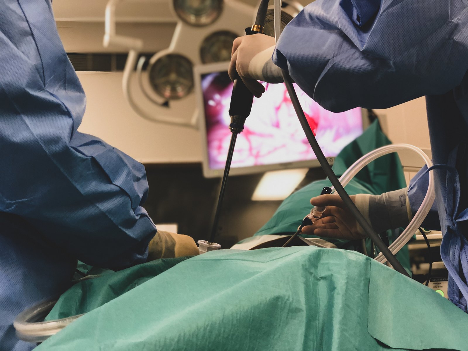
In certain cases, particularly in patients with locally advanced stomach cancer, diagnostic laparoscopy is performed before treatment begins. Through small incisions in the abdominal wall, the surgeon inserts a camera to examine the abdominal organs. This procedure can reveal small tumor deposits on the peritoneum that may not be visible on CT scans. If necessary, tissue or fluid samples are collected for further analysis.
5. Laboratory tests
Complete blood counts and blood chemistry tests help assess the patient's overall health, detect anemia, identify liver function abnormalities, and reveal other changes that may affect treatment. However, blood tests alone cannot confirm or rule out stomach cancer. Tumor markers such as CEA and CA 19-9 are also not reliable for the initial diagnosis of the disease.
6. Molecular and genetic testing of the tumor
After the diagnosis is confirmed, particularly in cases of advanced stomach cancer, additional tests may be performed on tumor tissue. Testing for HER2, PD-L1, MSI/MMR, and CLDN18.2 helps identify the tumor's biological characteristics and determine whether immunotherapy or targeted therapies may be suitable. The specific tests required depend on the stage of the disease and the planned treatment.
Based on the examination results, stomach cancer is staged according to the international TNM classification system, which considers the depth of tumor invasion, involvement of regional lymph nodes, and the presence of distant metastases. These findings, together with the tumor's histological and molecular characteristics, form the basis for developing an individualized treatment plan.
Stomach cancer treatment methods
Modern stomach cancer treatment is based on a multidisciplinary approach that may include endoscopic procedures, surgical removal of the tumor, chemotherapy, immunotherapy, targeted therapy, and radiation therapy. The choice of treatment depends on the stage of the disease, the location and size of the tumor, its histological and molecular characteristics, and the patient's overall health.
Surgical treatment of stomach cancer
Surgery remains the main curative treatment for stomach cancer and is used in most patients with localized tumors that can be completely removed. At the earliest stages, endoscopic removal of the tumor may be sufficient, allowing the stomach to be preserved. More advanced tumors require the removal of part or all of the stomach, together with regional lymph nodes. When the disease is diagnosed at a locally advanced stage, surgery is often part of a comprehensive treatment plan that also includes chemotherapy.
The choice of surgical technique depends on the size and location of the tumor, how deeply it has grown into the stomach wall, whether lymph nodes are affected, and the patient's general condition. Modern technologies allow many procedures to be performed using minimally invasive techniques, including robotic systems. However, the primary goal of any curative operation remains complete tumor removal while preserving the patient's quality of life as much as possible.
Endoscopic tumor resection (EMR and ESD)
Endoscopic tumor removal is used for early-stage stomach cancer when the malignant lesion is confined to the superficial layers of the stomach wall and the risk of lymph node involvement is extremely low. The procedure is performed using a flexible endoscope inserted through the mouth, without external incisions or removal of the stomach.
Two main techniques are used:
- Endoscopic mucosal resection (EMR) involves removing a small, superficial lesion using a special snare.
- Endoscopic submucosal dissection (ESD) is a more advanced technique that allows the tumor to be separated from the underlying tissue and removed in one piece, including relatively large superficial lesions.
The main advantage of endoscopic resection is that the stomach and its digestive function can be fully preserved. Recovery is usually faster than after conventional surgery. However, this method is suitable only for tumors with specific characteristics. After the procedure, the removed tissue is carefully examined to confirm that the tumor has been completely removed and that there are no features indicating a high risk of cancer spread.
At Helios Hospital Berlin-Buch, specialists in the Department of Gastroenterology perform endoscopic mucosal resection (EMR), endoscopic submucosal dissection (ESD), and endoscopic full-thickness resection for gastrointestinal tumors. The hospital has a large multidisciplinary endoscopy center where thousands of diagnostic and therapeutic procedures are performed every year.
Laparoscopic surgery
Laparoscopic gastrectomy is a minimally invasive technique for the surgical removal of the stomach. Instead of making one large incision, the surgeon creates several small openings in the abdominal wall through which a video camera and specialized instruments are inserted. The removed tissue is extracted through an additional small incision.
When clinically appropriate, both partial and total gastrectomy can be performed laparoscopically, including the necessary removal of lymph nodes. Compared with open surgery, this approach may reduce blood loss, postoperative pain, and the length of hospital stay, while also helping patients recover more quickly. However, the oncological completeness of the procedure must meet the same standards as open surgery.
Laparoscopic procedures are particularly beneficial for patients in whom the tumor can be safely removed using a minimally invasive approach. Open surgery may still be necessary in cases involving complex anatomy, extensive disease, or the need for major combined resections.
Da Vinci robotic surgery
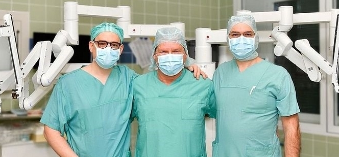
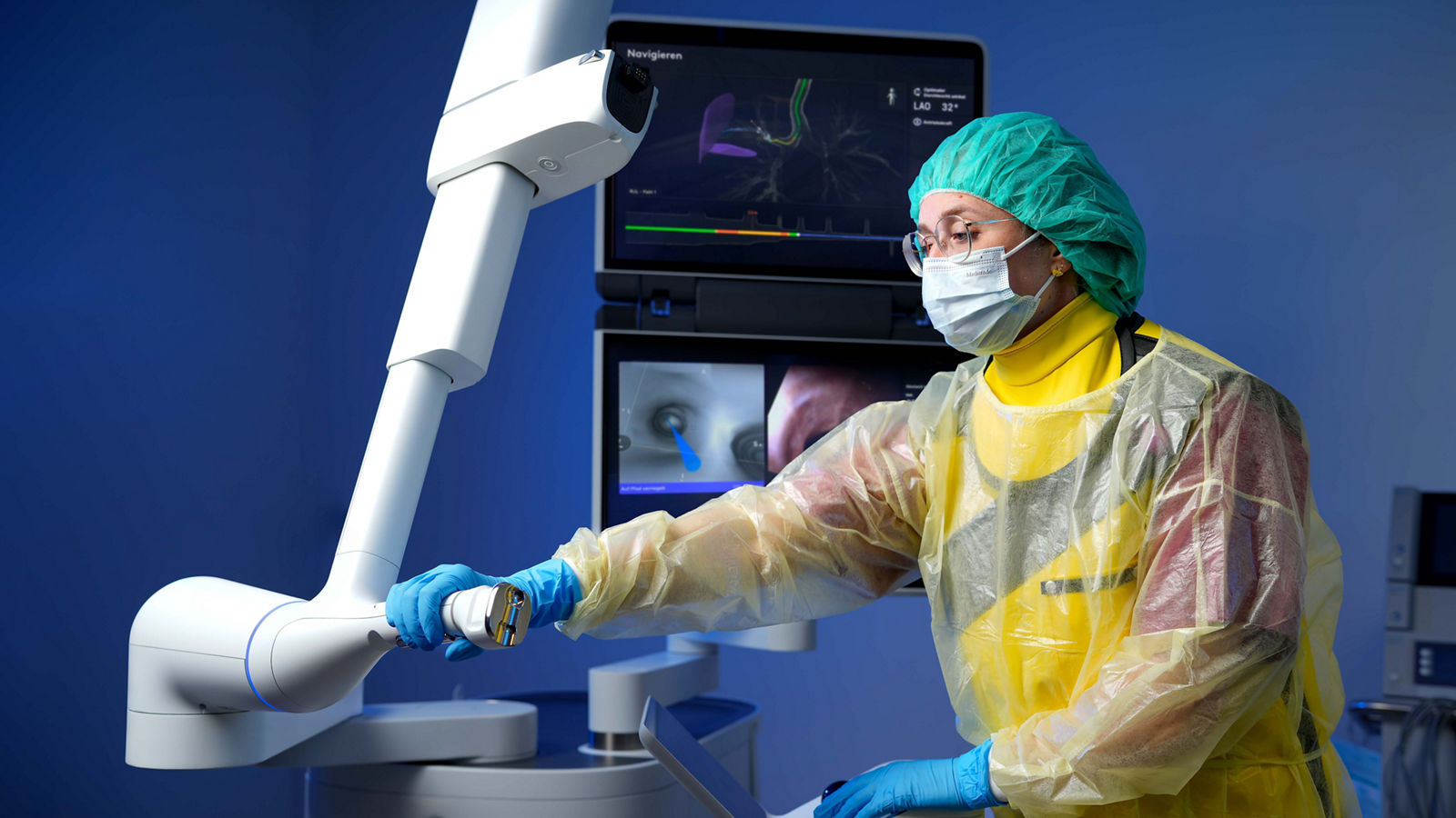
Robot-assisted surgery represents a further development of minimally invasive surgical techniques. When using the Da Vinci system, the surgeon controls miniature instruments from a specialized console while viewing the surgical field in a magnified three-dimensional image. The robot does not perform the operation independently; instead, it precisely reproduces the surgeon's movements.
The instruments' wide range of motion and detailed visualization are particularly useful during complex stages of gastrectomy, including dissection around major blood vessels, lymph node removal, and reconstruction of the digestive tract. The robotic approach allows these procedures to be performed through small incisions. In suitable patients, it may help reduce blood loss and accelerate recovery compared with open surgery. However, a clear long-term oncological advantage of robotic surgery over conventional laparoscopy has not yet been established.
One of the Helios centers specializing in these procedures is Helios Hospital Erfurt. The hospital is equipped with the Da Vinci Xi system and offers robot-assisted stomach surgery, including partial and total gastrectomy for malignant tumors. Its Department of General and Visceral Surgery is part of the Central Germany Robotic Surgery Center (Robotikzentrum Mitteldeutschland), which brings together specialized surgical teams and advanced robotic technologies.
At Helios Hospital Berlin-Buch, robot-assisted, laparoscopic, and open surgical techniques are also used to treat gastrointestinal tumors. The surgical approach is selected individually according to the complexity of the procedure and the patient's clinical circumstances.
Partial and total gastrectomy
When stomach cancer cannot be removed endoscopically, the main surgical treatment is gastrectomy — the removal of part or all of the stomach. The extent of surgery depends on the tumor's location and size, the depth of invasion, and whether all malignant tissue can be completely removed. Regional lymph nodes are usually removed together with the affected part of the stomach because they may contain cancer cells.
Partial (subtotal) gastrectomy involves removing a substantial portion of the stomach while preserving a healthy section. This procedure is most commonly performed for tumors located in the lower part of the stomach. After removing the affected tissue, the surgeon connects the remaining stomach to the small intestine, restoring continuity of the digestive tract. Because part of the stomach is preserved, patients can usually continue eating by mouth, although portion sizes become smaller and dietary habits need to be adjusted.
Total gastrectomy is a more extensive procedure in which the entire stomach is removed. It may be necessary for tumors in the upper part of the stomach, extensive involvement of the organ, or cases in which partial resection would not allow complete removal of the malignant tissue. After removing the stomach, the surgeon reconstructs the digestive tract by connecting the esophagus directly to the small intestine, most commonly using a Roux-en-Y reconstruction. This involves creating a loop of small intestine through which food passes from the esophagus into the intestine, where digestion and nutrient absorption continue.
Even after complete removal of the stomach, a person can eat independently by mouth and lead an active life. However, the absence of the stomach changes the digestive process: food is no longer stored or partially processed in the stomach but passes directly into the small intestine. Patients therefore need to eat small portions five or six times a day, or more frequently, chew food thoroughly, and gradually adapt their diet. Some patients may experience dumping syndrome after meals — a condition caused by food entering the intestine too quickly and associated with weakness, sweating, palpitations, nausea, or diarrhea.
After total gastrectomy, lifelong vitamin B12 supplementation is also necessary because the absence of the stomach prevents the production of intrinsic factor, which is required for vitamin B12 absorption. Doctors regularly monitor body weight, blood counts, iron levels, and other nutritional parameters. Additional nutritional support may be needed during the first few weeks after surgery, although most patients gradually return to eating independently.
An important component of curative surgical treatment is D2 lymphadenectomy — the systematic removal of specific groups of lymph nodes around the stomach and along major blood vessels. This procedure helps remove tissue that may contain cancer cells and allows more accurate staging of the disease. Depending on the clinical situation, gastrectomy may be performed using an open, laparoscopic, or robot-assisted approach.
Chemotherapy for stomach cancer
Chemotherapy plays an important role in the comprehensive treatment of stomach cancer. It is used both before and after surgical removal of the tumor, as well as in advanced disease when curative surgery is not possible. Anticancer drugs target rapidly dividing cells, helping to reduce tumor size, destroy microscopic cancer deposits, and slow disease progression.
The effectiveness of chemotherapy for stomach cancer has improved considerably in recent years. This progress is due not only to the development of new drugs but also to more effective drug combinations, better treatment sequencing, and improved management of side effects. For example, the FLOT4 clinical trial showed that the modern FLOT regimen, administered before and after surgery, increased median overall survival to 50 months compared with 35 months for the previously widely used ECF/ECX regimens. However, results from individual studies cannot be directly applied to every patient: treatment effectiveness depends on the stage of the disease, tumor characteristics, and the patient's overall health.
Depending on its purpose, chemotherapy may be used in several ways:
- Neoadjuvant chemotherapy — before surgery. Helps shrink the tumor, target potential micrometastases, and increase the likelihood of completely removing malignant tissue.
- Adjuvant chemotherapy — after surgery. Aims to destroy any remaining microscopic cancer cells and reduce the risk of recurrence.
- Perioperative chemotherapy — before and after surgery. Combines both approaches and is an important part of treatment for many patients with resectable, locally advanced stomach cancer.
- Palliative chemotherapy. Used for unresectable or metastatic disease to control tumor growth, relieve symptoms, and prolong survival.
One of the most extensively studied and widely used regimens for resectable stomach cancer is FLOT, which combines four drugs: 5-fluorouracil, leucovorin (folinic acid), oxaliplatin, and docetaxel. Treatment typically consists of four two-week cycles before surgery and four cycles afterward, provided the patient's condition allows treatment to continue. For patients who are not suitable for this intensive four-drug regimen, doctors may select less intensive combinations, such as FOLFOX (5-fluorouracil, leucovorin, and oxaliplatin) or CAPOX (capecitabine and oxaliplatin), depending on the clinical circumstances.
For metastatic stomach cancer, combinations of fluoropyrimidines and platinum-based drugs are commonly used. Paclitaxel, irinotecan, and other cytotoxic drugs may be introduced in subsequent lines of treatment. In recent years, treatment options have expanded through the combination of chemotherapy with immunotherapy and targeted drugs. The choice of these combinations depends, among other factors, on the tumor's molecular characteristics, including HER2, PD-L1, MSI/MMR, and CLDN18.2 status.
Despite improvements in effectiveness, chemotherapy may cause nausea, fatigue, low blood cell counts, increased susceptibility to infections, and peripheral neuropathy — a condition affecting sensation in the hands and feet. Modern supportive care, including antiemetic medications, dose adjustments, complication prevention, and nutritional support, helps many patients tolerate treatment better. Monitoring nutrition and body weight is particularly important because stomach cancer is often associated with loss of appetite and weight loss.
At Helios University Hospital Wuppertal, stomach cancer treatment is organized within a specialized center for esophageal and gastric surgery. During multidisciplinary tumor board meetings, specialists determine whether preoperative chemotherapy is needed, assess the feasibility of surgery, and develop an individualized treatment sequence. Helios Hospital München West also takes a comprehensive approach to treating gastrointestinal tumors, involving oncologists, surgeons, and other specialists. Systemic treatment plans are discussed at regular multidisciplinary tumor board meetings.
Local intraperitoneal chemotherapy: HIPEC and PIPAC
In advanced stomach cancer, malignant cells may spread to the peritoneum — the thin membrane lining the abdominal cavity and covering the organs within it. This condition is known as peritoneal metastasis or peritoneal carcinomatosis. Treating it is particularly challenging because systemic chemotherapy does not always provide sufficient control of tumor deposits in this area.
For selected patients, local intraperitoneal chemotherapy techniques known as HIPEC and PIPAC have been developed. Unlike conventional chemotherapy, these methods deliver anticancer drugs directly into the abdominal cavity, allowing high local concentrations of medication to be achieved. Both techniques are performed in specialized centers, but they differ in how the drugs are administered, whether surgery is required, and which patients may be eligible. In stomach cancer, they do not replace standard systemic treatment, and their potential benefits must be assessed individually.
Hyperthermic intraperitoneal chemotherapy (HIPEC)
HIPEC (Hyperthermic Intraperitoneal Chemotherapy) involves delivering a heated chemotherapy solution directly into the abdominal cavity. It is usually performed in combination with cytoreductive surgery (CRS), during which the surgeon removes all visible tumor deposits from the peritoneum and, when necessary, affected areas of other organs.
After the surgical stage, the abdominal cavity is perfused with a chemotherapy solution heated to approximately 41–43°C. A specialized circulation system ensures that the solution is distributed evenly. The procedure usually takes between 60 and 120 minutes. Elevated temperatures may enhance the effects of certain cytotoxic drugs on cancer cells, while local administration allows treatment to target microscopic tumor deposits that cannot be identified or removed during surgery.
The main advantage of HIPEC is the opportunity to combine the most complete possible surgical removal of visible metastases with direct chemotherapy exposure to any remaining cancer cells in the abdominal cavity. This approach may be considered for patients with limited peritoneal involvement, a satisfactory general condition, and a high likelihood of complete removal of visible tumor deposits.
However, CRS and HIPEC constitute a complex combined treatment that may involve serious postoperative complications and a lengthy recovery period. Furthermore, in stomach cancer, the additional overall survival benefit of HIPEC has not yet been demonstrated convincingly enough. The technique is therefore not part of routine standard treatment and should be considered in specialized centers, preferably within clinical trials.
At Helios Dr. Horst Schmidt Hospital Wiesbaden, a specialized cytoreductive surgery and HIPEC program is available for patients with peritoneal tumor involvement, including metastatic stomach cancer. Before treatment, specialists assess the extent of the disease, the feasibility of complete tumor removal, and surgical risks. Decisions are made by a multidisciplinary tumor board, and procedures are performed by a team that includes experienced visceral surgeons, oncologists, anesthesiologists, and intensive care specialists.
Pressurized intraperitoneal aerosol chemotherapy (PIPAC)
PIPAC (Pressurized Intraperitoneal Aerosol Chemotherapy) is a newer technique in which chemotherapy is delivered into the abdominal cavity as a fine aerosol under pressure. It was developed to target peritoneal metastases locally, including situations in which complete surgical removal of tumor deposits is not possible.
The procedure is performed laparoscopically under general anesthesia. Through small incisions in the abdominal wall, the doctor inserts a camera and a specialized aerosol delivery device. Once the required pressure has been established, the chemotherapy drug is distributed throughout the abdominal cavity as an aerosol and remains in contact with the affected tissues for a specified period, usually around 30 minutes. The aerosol is then removed through a closed filtration system.
Unlike HIPEC, PIPAC generally does not require the prior removal of all visible tumor deposits. The procedure can be repeated several times at defined intervals, allowing doctors to assess the tumor's response to treatment. When appropriate, it may be combined with systemic chemotherapy.
The potential advantages of PIPAC include minimally invasive drug delivery, the possibility of repeated procedures, and direct exposure of peritoneal metastases to chemotherapy. Local administration may reduce systemic exposure to certain chemotherapy drugs, although this does not mean that side effects are absent.
PIPAC is primarily considered for patients with unresectable peritoneal metastases when standard systemic treatment does not provide adequate disease control or when combined treatment approaches are being investigated. Despite encouraging findings from individual studies, there is still insufficient convincing evidence that PIPAC prolongs overall survival in stomach cancer. For this reason, its use for this disease in Germany is recommended only within clinical trials.
At Helios Hospital Schwerin, both HIPEC and PIPAC technologies are available. In an official hospital publication, specialists describe the use of PIPAC in patients with peritoneal metastases, including those originating from stomach cancer. The procedure is performed laparoscopically and may complement systemic chemotherapy. The hospital emphasizes the importance of careful patient selection and the investigational status of the technique.
Immunotherapy for stomach cancer
Immunotherapy has become one of the most significant advances in stomach cancer treatment in recent years. Unlike chemotherapy, which directly targets rapidly dividing cells, immunotherapy drugs help the patient's own immune system recognize and attack malignant tumors. Today, immunotherapy is used not only for metastatic stomach cancer but also in combination with chemotherapy and surgery for certain resectable tumors.
The main approach in modern immunotherapy involves immune checkpoint inhibitors. Cancer cells can exploit proteins such as PD-1 and PD-L1 to suppress immune cell activity and avoid destruction. Specialized monoclonal antibodies block these mechanisms, restoring the immune system's ability to fight the disease. However, immunotherapy is not suitable for every patient: its effectiveness depends largely on the tumor's molecular characteristics, the stage of the disease, and the selected drug combination.
Several types of immunotherapy are used for stomach cancer in Germany:
- Nivolumab (Opdivo) is a PD-1 inhibitor used in combination with chemotherapy for certain forms of advanced or metastatic HER2-negative stomach cancer. In Europe, this treatment is approved for tumors with a PD-L1 combined positive score (CPS) of 5 or higher. The CheckMate 649 trial showed that adding nivolumab to chemotherapy improved overall survival compared with chemotherapy alone.
- Pembrolizumab (Keytruda) is another PD-1 inhibitor. It may be administered with chemotherapy for advanced HER2-negative stomach cancer with a PD-L1 CPS of 1 or higher. For HER2-positive tumors with the appropriate PD-L1 expression, pembrolizumab may be combined with trastuzumab and chemotherapy. Tumors with high microsatellite instability (MSI-H) or deficient DNA mismatch repair (dMMR) may be particularly sensitive to PD-1 inhibitors.
- Tislelizumab (Tevimbra) is a PD-1 inhibitor used in combination with chemotherapy as first-line treatment for certain HER2-negative advanced or metastatic gastric adenocarcinomas. In Europe, eligibility depends on PD-L1 expression measured using a tumor area positivity (TAP) score of 5% or higher.
- Durvalumab (Imfinzi) is a PD-L1 inhibitor approved in the European Union since 2026 for perioperative treatment of resectable gastric and gastroesophageal junction adenocarcinoma. The drug is administered together with FLOT chemotherapy before and after surgery, followed by durvalumab alone.
How effective is modern immunotherapy?
The effectiveness of immunotherapy has been demonstrated in major international clinical trials. In advanced stomach cancer, adding PD-1 inhibitors to standard chemotherapy can prolong survival and improve disease control in certain patient groups. For example, in the CheckMate 649 trial, median overall survival among patients with a PD-L1 CPS of 5 or higher was 14.4 months with nivolumab plus chemotherapy, compared with 11.1 months with chemotherapy alone. These figures represent the results of a specific group of trial participants and should not be interpreted as an individual patient's prognosis.
An especially important development has been the expansion of immunotherapy into the treatment of resectable stomach cancer. In the phase III MATTERHORN trial, combining durvalumab with FLOT before and after surgery increased the two-year event-free survival rate — meaning survival without events such as disease progression, recurrence, or death — to 67.4%, compared with 58.5% for FLOT without durvalumab. Subsequent results also confirmed a statistically significant improvement in overall survival. Based on these findings, the combination received European Union approval in March 2026.
Before immunotherapy is prescribed, the tumor undergoes molecular and pathological testing. Doctors assess PD-L1 expression, MSI/MMR status, and HER2 status, while also considering the patient's general condition, coexisting diseases, and previous treatments. It is important to remember that immunotherapy may cause specific side effects related to excessive immune system activity, including inflammation of the intestines, lungs, liver, and endocrine organs. Treatment therefore requires regular medical monitoring.
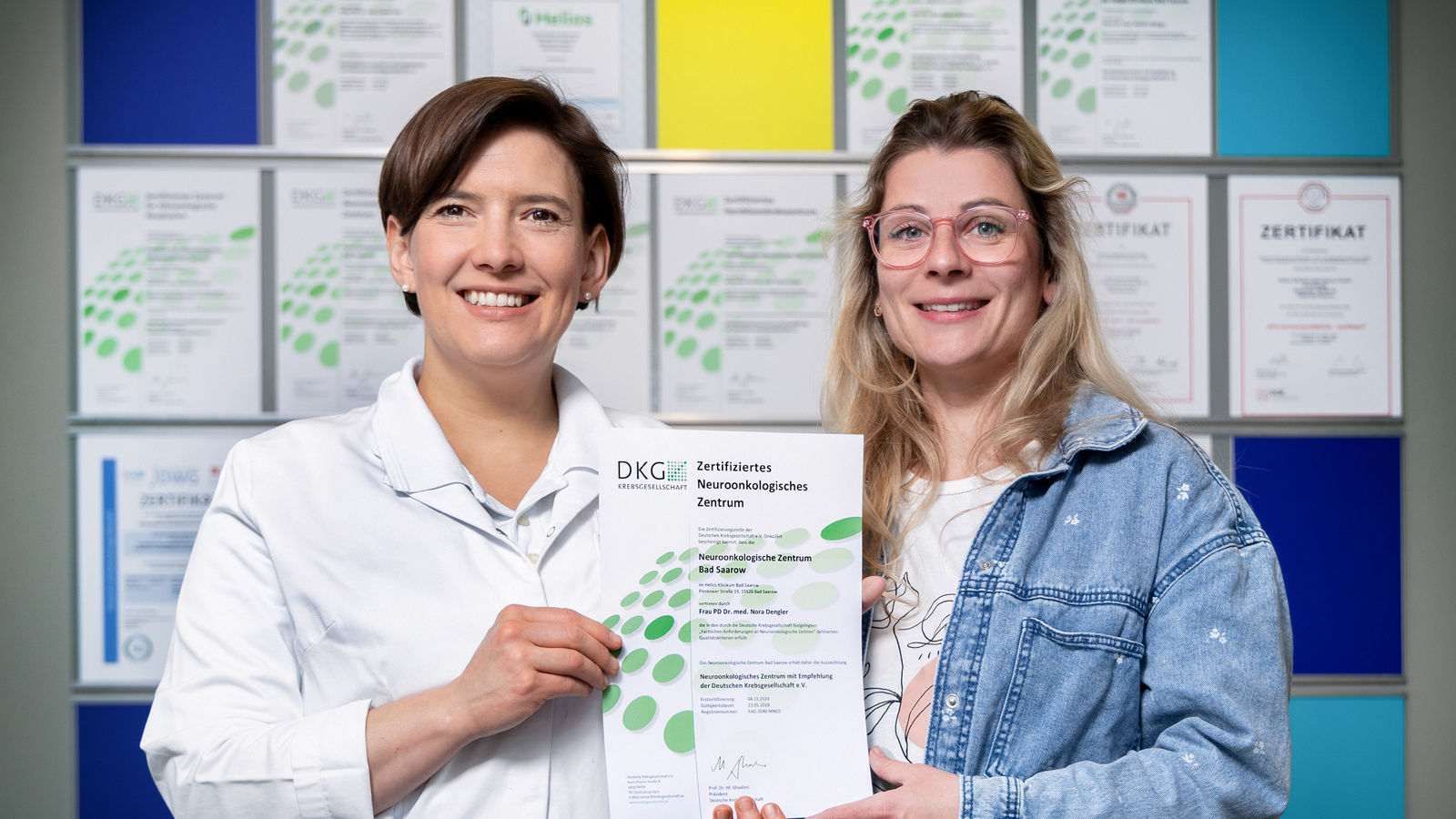
At Helios Hospital Bad Saarow, modern immunotherapy approaches for stomach cancer are being investigated in international clinical trials. In particular, the oncology center participated in the DANTE/FLOT8 trial, which investigated the combination of perioperative FLOT chemotherapy with the PD-L1 inhibitor atezolizumab. The hospital's official website also lists the ARTEMIDE-Gastric01 trial, which investigates a new immunotherapy combination for patients with HER2-positive advanced stomach cancer.
At Helios Hospital Emil von Behring in Berlin, the Department of Hematology and Oncology offers treatment with monoclonal antibodies and participation in clinical trials. The suitability of a particular immunotherapy regimen for stomach cancer is assessed individually after evaluating the tumor's molecular characteristics and discussing the case at a multidisciplinary tumor board.
Targeted therapy for stomach cancer
Targeted therapy is one of the most promising approaches in personalized stomach cancer treatment. Unlike conventional chemotherapy, which primarily affects rapidly dividing cells, targeted drugs act on specific molecules and signaling mechanisms involved in tumor growth and spread. This makes it possible to select treatment based on the biological characteristics of cancer cells rather than the stage of the disease alone.
The range of targeted therapies has expanded considerably in recent years. While treatment initially focused mainly on the HER2 protein, current options also include drugs that block the formation of new tumor blood vessels and newer antibodies directed against the Claudin 18.2 protein. However, targeted treatment is not suitable for every patient. Molecular and pathological testing of tumor tissue is necessary before treatment to identify the relevant targets.
The main types of targeted therapy used for stomach cancer include:
-
HER2-targeted therapy — trastuzumab (Herceptin). Approximately 15–20% of gastric and gastroesophageal junction adenocarcinomas show increased expression of the HER2 protein, which promotes cancer cell growth. Trastuzumab is a monoclonal antibody that blocks this receptor. In HER2-positive advanced stomach cancer, it is usually administered with chemotherapy and, for certain indications, with the immunotherapy drug pembrolizumab. The advantage of this approach is its ability to specifically target tumors whose growth is associated with HER2.
-
Antibody-drug conjugates — trastuzumab deruxtecan (Enhertu). This is a more advanced form of targeted therapy that combines a monoclonal antibody against HER2 with a potent cytotoxic drug. The antibody recognizes HER2-expressing cancer cells, allowing the drug component to be delivered preferentially to them. This mechanism combines targeted action with the anticancer effects of chemotherapy. Trastuzumab deruxtecan is used for certain forms of HER2-positive advanced stomach cancer following previous treatment with trastuzumab. One important risk is interstitial lung disease, making careful monitoring essential.
-
Antiangiogenic therapy — ramucirumab (Cyramza). Malignant tumors need to develop new blood vessels in order to grow. Ramucirumab blocks VEGFR-2, a receptor involved in this process, thereby interfering with the development of the tumor's blood supply. The drug is mainly used for advanced stomach cancer after disease progression following previous chemotherapy, often in combination with paclitaxel. Unlike HER2-targeted treatment, ramucirumab does not require confirmation of a specific mutation or increased HER2 expression.
-
CLDN18.2-targeted therapy — zolbetuximab (Vyloy). One of the newer approaches to stomach cancer treatment targets Claudin 18.2, a protein found on the surface of cells in certain tumors. Zolbetuximab is a monoclonal antibody that recognizes this protein and activates immune mechanisms that destroy cancer cells. In the European Union, the drug is approved in combination with chemotherapy as first-line treatment for certain HER2-negative, CLDN18.2-positive, locally advanced unresectable or metastatic gastric and gastroesophageal junction adenocarcinomas. CLDN18.2 expression must be confirmed by specialized immunohistochemical testing before treatment.
How effective is targeted therapy?
The effectiveness of modern targeted drugs has been demonstrated in major clinical trials. For example, the ToGA trial showed that adding trastuzumab to chemotherapy increased median overall survival in patients with HER2-positive advanced stomach cancer from 11.1 to 13.8 months. In the RAINBOW trial, ramucirumab plus paclitaxel increased median overall survival from 7.4 to 9.6 months compared with paclitaxel alone in previously treated patients.
The results of the SPOTLIGHT and GLOW trials investigating zolbetuximab are particularly important. They demonstrated improvements in both progression-free survival and overall survival when the drug was added to standard chemotherapy in patients with CLDN18.2-positive tumors. These findings confirm that molecular testing can identify patient groups for whom a particular treatment may be especially effective.
The main advantage of targeted therapy is the ability to individualize treatment according to the tumor's molecular characteristics. However, targeted action does not mean that these drugs are free from side effects. Depending on the medication, patients may experience nausea, diarrhea, changes in blood counts, increased blood pressure, cardiovascular complications, and other adverse reactions. Like chemotherapy, targeted treatment therefore requires regular monitoring by an oncologist.
At Helios Hospital Bad Saarow, new approaches to personalized stomach cancer treatment are being investigated. In particular, the hospital lists participation in the phase III ARTEMIDE-Gastric01 trial, which is studying the combination of trastuzumab deruxtecan with the investigational immunotherapy drug rilvegostomig and a fluoropyrimidine in patients with HER2-positive advanced stomach cancer. The hospital also lists the FRUQUITAS trial, which investigates the angiogenesis inhibitor fruquintinib in combination with trifluridine/tipiracil for metastatic disease.
Radiation therapy for stomach cancer
Radiation therapy (radiotherapy) uses high-energy ionizing radiation to damage the DNA of cancer cells, causing them to lose their ability to divide and eventually die. In stomach cancer, radiation therapy is used much less frequently than surgery or chemotherapy. This is partly because the stomach constantly changes its position and shape, while radiation-sensitive organs — including the liver, kidneys, intestines, and spinal cord — are located nearby.
In modern oncology, radiation therapy is not a routine component of treatment for most resectable stomach tumors. Curative treatment is generally based on surgery and perioperative systemic therapy. However, in certain clinical situations, radiation can help control the tumor, reduce the risk of local recurrence, or relieve symptoms. Advances in high-precision technologies have made radiation therapy more targeted and improved protection of surrounding healthy tissues.
Depending on the stage of the disease and treatment goals, radiation therapy may be used in the following situations:
- Postoperative chemoradiotherapy. In selected cases, radiation is combined with chemotherapy after surgery, for example when the tumor could not be completely removed and microscopic examination reveals malignant cells at the resection margin (R1). This approach may improve local disease control but is not routinely used after a properly performed curative operation.
- Treatment of unresectable localized tumors. If the cancer has not spread to distant organs but surgery is impossible because of the extent of the tumor or the patient's condition, chemoradiotherapy may be considered. The aim is to achieve the longest possible disease control and, in selected cases, to pursue treatment with curative intent.
- Palliative radiation therapy. Used in advanced stomach cancer to reduce tumor-related bleeding, pain, difficulty with food passage, and other severe symptoms. Shorter courses of radiation are generally used in these situations to reduce the burden of treatment.
- Radiation therapy for individual metastases. In certain situations involving a limited number of metastatic lesions, highly precise local radiation therapy may be considered. However, systemic drug treatment remains the main approach for metastatic stomach cancer, and decisions about local radiation are made individually.
Modern high-precision radiation therapy techniques
One of the major advances in radiation oncology has been the introduction of technologies that allow doctors to calculate radiation doses precisely and adapt their distribution to the shape of the tumor. This is particularly important in stomach cancer because of the proximity of vital organs.
Intensity-modulated radiation therapy (IMRT) allows the intensity of radiation to be adjusted across different parts of the treatment area. This enables doctors to create complex dose distributions that cover the tumor while reducing exposure to surrounding healthy tissues. The technique is particularly useful when a tumor is located close to sensitive organs.
Image-guided radiation therapy (IGRT) uses X-ray imaging or computed tomography immediately before treatment and, when necessary, during radiation delivery. This allows the position of the target area to be checked and the patient's positioning to be adjusted. Because the stomach's position depends on breathing and how full it is, image guidance is especially important for treatment accuracy.
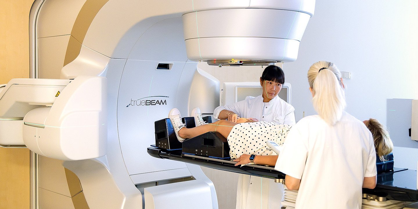
Tomotherapy combines the principles of IMRT and computed tomography. During treatment, the radiation source rotates around the patient, delivering radiation from different directions. This technology allows complex treatment plans to be created while monitoring the position of anatomical structures. It can be useful when treating large or anatomically complex areas.
Stereotactic body radiation therapy (SBRT) delivers high doses of radiation to a small, precisely defined target over a limited number of treatment sessions. In stomach cancer, this method may be considered for treating individual metastases, for example in the liver or lungs, in carefully selected patients. However, it is not a standard method for irradiating the primary stomach tumor.
Modern treatment planning techniques help reduce unnecessary radiation exposure to healthy tissues, but they do not eliminate side effects. Patients may experience nausea, loss of appetite, fatigue, irritation of mucous membranes, and digestive disturbances during treatment. Radiation therapy plans are therefore developed individually, taking into account the tumor's location, previous treatment, and organ function.
At Helios Hospital Berlin-Buch, a specialized Department of Radiation Therapy and Radiation Oncology provides both curative and palliative radiation treatment. The hospital's oncology center offers advanced technologies, including tomotherapy, high-precision image-guided radiation therapy, and combined chemoradiotherapy. Specialists also provide radiation treatment for metastatic lesions. In stomach cancer, the need for radiation therapy is determined by a multidisciplinary tumor board involving oncologists, surgeons, and radiation oncologists.
Life after stomach cancer treatment
After completing the main course of stomach cancer treatment, patients enter an important phase of recovery and adjustment to everyday life. Modern surgical techniques, systemic therapies, and rehabilitation programs allow many patients to gradually regain their physical strength, return to work, travel, and enjoy their usual activities. The duration and nature of recovery depend on the treatment received, the patient's overall health, and whether part or all of the stomach has been removed.
Proper nutrition, a gradual increase in physical activity, regular medical follow-up, and timely correction of nutritional deficiencies are particularly important during this period. Rehabilitation programs are tailored to each patient's needs, with the main goal of helping them regain strength, adapt to changes in digestion, and maintain a good quality of life.
Rehabilitation after surgery and cancer treatment
Recovery after surgery begins while the patient is still in the hospital. Under medical supervision, patients gradually resume walking, breathing exercises, and eating. Early mobilization helps maintain muscle strength, improve bowel function, and reduce the risk of postoperative complications.
After discharge, rehabilitation may include therapeutic exercises, moderate aerobic activity, physiotherapy, and consultations with a dietitian. Physical activity is increased gradually: patients typically begin with short walks before progressing to longer walks, swimming, cycling, and other activities approved by their doctor. Following chemotherapy or radiation therapy, additional attention is given to restoring stamina, muscle mass, and overall well-being.
In Germany, specialized oncology rehabilitation programs (onkologische Rehabilitation) are available to patients following cancer treatment. These programs combine physical therapy, nutritional guidance, psychological support, and assistance with returning to everyday and professional activities. Gastroenterologists, oncologists, clinical nutrition specialists, and psycho-oncologists may also be involved in the recovery process when needed.
Nutrition after partial or total gastrectomy
After gastrectomy, the body needs time to adapt to changes in the digestive process. Even after complete removal of the stomach, patients can eat independently, although their usual eating habits need to be adjusted.
Patients are generally advised to eat small meals five or six times a day, or more frequently, chew food thoroughly, and introduce new foods gradually. Adequate protein and calorie intake is essential to maintain body weight and support tissue recovery. If necessary, a doctor or dietitian may recommend specialized nutritional supplements.
Some patients experience dumping syndrome after surgery, a condition in which food passes too quickly into the small intestine. To reduce symptoms, patients are generally advised to avoid large meals, limit foods high in rapidly absorbed sugars, and, when appropriate, drink most fluids between meals rather than during meals.
After total gastrectomy, lifelong vitamin B12 supplementation is necessary. Doctors may also prescribe iron, calcium, vitamin D, and other nutrients depending on blood test results. Regular monitoring of body weight and blood parameters helps identify nutritional deficiencies early and allows dietary adjustments to be made promptly.
Medical follow-up: Which examinations are needed and how often?
After stomach cancer treatment, each patient receives an individualized follow-up care plan. Its main purposes are to monitor recovery, assess nutritional status and general health, identify changes requiring medical attention, and support long-term quality of life.
The frequency of follow-up visits depends on the stage of the disease, the extent of surgery, the systemic treatment received, and individual risk factors. Appointments are generally more frequent during the first few years after treatment, with longer intervals possible later.
The main components of follow-up care include:
- Oncologist or gastroenterologist consultations. The doctor assesses the patient's well-being, body weight, food tolerance, digestive function, and recovery after treatment. An indicative schedule may involve appointments every 3–6 months during the first two years, followed by visits every 6–12 months until the fifth year. The exact schedule is determined individually.
- Laboratory tests. Depending on the surgery performed, doctors monitor complete blood counts, hemoglobin, iron and ferritin levels, vitamin B12, and, when necessary, folic acid, vitamin D, calcium, and other nutritional markers. Lifelong monitoring of nutritional status is particularly important after total gastrectomy.
- Gastroscopy. After partial gastrectomy or endoscopic treatment, the doctor may recommend follow-up gastroscopy to examine the lining of the remaining stomach. Following endoscopic resection of early stomach cancer, regular endoscopic surveillance is generally required, often annually, depending on histological findings and the condition of the gastric mucosa. Routine gastroscopy is not necessary for every patient after total gastrectomy.
- Computed tomography (CT) and other imaging examinations. CT scans of the chest, abdomen, and pelvis, ultrasound examinations, or other imaging techniques may be recommended when clinically indicated. The need for and frequency of these examinations are determined individually; there is no universal schedule for routine CT scans that applies to all patients after curative stomach cancer treatment.
If new persistent symptoms, difficulties with eating, or unexplained changes in body weight occur, patients should contact their doctor rather than wait for the next scheduled appointment.
What should patients avoid or limit after treatment?
Recovery from stomach cancer does not usually require lifelong restrictions on physical activity, travel, or everyday activities. However, certain precautions can help the body adapt to treatment and maintain good health.
During the first few weeks after surgery, patients should avoid heavy lifting and strenuous physical activity until their surgeon confirms that these activities are safe. Once recovery progresses, physical activity is not only permitted but encouraged, provided that its intensity is increased gradually.
Patients are advised to stop smoking completely and avoid alcohol whenever possible, particularly during recovery and systemic cancer treatment. After gastrectomy, overeating, long intervals between meals, and excessive consumption of sugary drinks or foods that may trigger dumping syndrome should be avoided. However, a universally restrictive diet is not necessary: nutritional recommendations are tailored to each patient's individual food tolerance.
Patients should also avoid taking high-dose vitamins, dietary supplements, or products marketed as "immune boosters" without consulting their doctor, as these may interact with prescribed treatments.
Long-term outlook and quality of life after treatment
When stomach cancer is detected early and treated appropriately, there is a strong possibility of achieving long-term disease control and complete recovery. Particularly favorable outcomes can be achieved when tumors are completely removed at an early stage, including through organ-preserving endoscopic procedures.
Modern surgical technologies, personalized systemic therapies, and rehabilitation programs help patients not only complete their treatment but also return to fulfilling everyday lives. Even after total gastrectomy, many people gradually adapt to a new eating routine, remain physically active, work, and travel.
Regular follow-up care, balanced nutrition, physical activity, and ongoing professional support help patients maintain the benefits of treatment and enjoy a good quality of life for many years.
Stomach cancer treatment at Helios Hospitals
Stomach cancer treatment at Helios Hospitals in Germany combines advanced medical technologies, the expertise of specialized oncology teams, and a personalized approach to every patient. With an extensive network of multidisciplinary hospitals and specialized centers, patients can receive comprehensive care — from confirming the diagnosis and determining the stage of the disease to surgery, systemic treatment, rehabilitation, and long-term follow-up.
One of the key advantages of Helios is its multidisciplinary approach to cancer treatment. Gastroenterologists, oncologists, visceral surgeons, radiologists, pathologists, and radiation oncologists work together to develop treatment strategies. At multidisciplinary tumor board meetings, specialists jointly review diagnostic findings and determine the most appropriate sequence of treatment. Their decisions take into account not only the stage and location of the tumor but also its molecular characteristics, the patient's coexisting medical conditions, and individual needs.
Particular emphasis is placed on accurate diagnosis. Specialized Helios centers use advanced endoscopic systems, endoscopic ultrasound, computed tomography (CT), magnetic resonance imaging (MRI), and molecular pathology testing of tumor tissue. Identifying biomarkers such as HER2, PD-L1, MSI/MMR, and CLDN18.2 helps doctors assess whether patients may benefit from modern targeted therapies and immunotherapy.
The advanced technological infrastructure of Helios Hospitals supports a wide range of stomach cancer treatment options. Depending on medical indications and the capabilities of the specialized center, patients may have access to organ-preserving endoscopic procedures, laparoscopic surgery, and robot-assisted operations, including procedures performed using the Da Vinci surgical system. Comprehensive treatment options also include modern chemotherapy regimens, immunotherapy, targeted therapy, and high-precision radiation therapy. For selected patients with advanced disease, specialized centers may consider additional approaches, including hyperthermic intraperitoneal chemotherapy (HIPEC). The suitability of each technique is assessed individually.
The professional expertise of the medical teams is another important advantage. Helios physicians collaborate closely with colleagues across different specialties, regularly update their professional skills, and follow modern clinical guidelines. Some oncology centers within the network participate in international clinical trials aimed at improving surgical techniques and developing new combinations of anticancer drugs. For patients who meet the eligibility criteria, these studies may provide access to additional treatment options.
The personalized approach at Helios extends beyond medical decisions to the organization of the patient's entire hospital stay. International patients can receive assistance with treatment preparation, the coordination of consultations and diagnostic examinations, and support throughout the different stages of their medical program. International patient services help manage organizational matters and facilitate communication with the medical team in a language the patient understands.
Patient comfort during hospitalization is also a priority. Modern patient rooms, options for upgraded accommodation, attentive medical staff, and assistance with everyday needs help create a calm and supportive environment, which is especially important during prolonged cancer treatment. When needed, care programs may also include dietary consultations, psychological support, and rehabilitation services.
Ultimately, stomach cancer treatment at Helios is centered on each patient's individual needs. The combination of precise diagnostics, advanced treatment methods, experienced multidisciplinary teams, and coordinated patient support enables a comprehensive and personalized approach — from the initial consultation through recovery after treatment.
If you are interested in arranging diagnosis or treatment at one of the Helios hospitals, you can submit a request using the contact form on our website or contact us via the email address provided at the top of the website.
Learn more about arranging treatment at Helios hospitals ⟶
Do you need more information about Helios Hospitals or want to schedule your treatment?