
Esophageal cancer: Effective treatment options
- Risk factors and causes
- Symptoms
- Diagnosis
- Esophageal cancer treatment methods
- Rehabilitation after esophageal resection
- Esophageal cancer treatment at Helios Hospitals
There are two main histological types of esophageal cancer: squamous cell carcinoma and adenocarcinoma. Squamous cell carcinoma develops from squamous epithelial cells and can occur in different parts of the esophagus. Adenocarcinoma arises from glandular cells and is most commonly found in the lower part of the esophagus, near the junction with the stomach. In Western countries, including Germany, adenocarcinoma is now the most common type of esophageal cancer.
In its early stages, the tumor may cause no characteristic symptoms for a long time. As it grows, it can invade the wall of the esophagus, spread to surrounding tissues and lymph nodes, and, at advanced stages, form distant metastases. The stage of the disease and the histological type of the tumor are key factors in determining the most appropriate treatment strategy.
Esophageal cancer usually develops not as a result of a single cause, but through a combination of factors that damage the esophageal lining over time or contribute to abnormal cellular changes. Importantly, the risk factors for squamous cell carcinoma and adenocarcinoma differ considerably.
Risk factors and causes of esophageal cancer
Esophageal cancer develops as a result of accumulated changes in the cells lining the esophagus, causing them to divide uncontrollably and become malignant. In most cases, it is not possible to identify a single cause of the disease in an individual patient. Importantly, the risk factors differ considerably between the two main types of esophageal cancer — squamous cell carcinoma and adenocarcinoma.
The most important risk factors include:
- Smoking and alcohol consumption — particularly important risk factors for esophageal squamous cell carcinoma. Their combined effect increases the risk more than either factor alone.
- Gastroesophageal reflux disease (GERD) — long-term exposure of the esophagus to acidic stomach contents can repeatedly damage its lining. Chronic reflux is primarily associated with an increased risk of adenocarcinoma.
- Barrett’s esophagus — one of the most important precancerous conditions. Long-term reflux can cause the normal cells lining the lower esophagus to be replaced by a different type of cell. Most people with Barrett’s esophagus do not develop cancer, but their risk of adenocarcinoma is increased, particularly when dysplasia is present.
- Esophageal achalasia — a condition in which the lower esophageal sphincter does not relax properly, interfering with the passage of food into the stomach. Long-term food retention and chronic inflammation of the esophageal lining increase the risk of cancer, particularly squamous cell carcinoma.
- Overweight and obesity — are associated with an increased risk of esophageal adenocarcinoma. One possible contributing mechanism is the higher likelihood of chronic gastroesophageal reflux.
- Previous damage and diseases of the esophagus — the risk may be increased after severe chemical burns of the esophagus that result in scar tissue and narrowing, as well as in certain rare conditions associated with chronic damage to the esophageal lining.
- Age and sex — esophageal cancer is considerably more common in older adults and in men.
- Diet and lifestyle — a low intake of fruit and vegetables and certain dietary patterns may be associated with an increased risk. Regular consumption of very hot beverages is also considered a risk factor for squamous cell carcinoma because of repeated thermal injury to the esophageal lining.
- Rare inherited disorders — for example, tylosis (an inherited condition characterized by thickening of the skin on the palms and soles) is associated with a substantially increased risk of esophageal squamous cell carcinoma. However, inherited syndromes account for only a small proportion of cases.
Among pre-existing conditions, chronic gastroesophageal reflux disease, Barrett’s esophagus, and achalasia are particularly important. Their roles differ: GERD and Barrett’s esophagus are primarily associated with the development of adenocarcinoma, whereas achalasia is more strongly associated with squamous cell carcinoma.
Having a risk factor or a precancerous condition does not mean that cancer will necessarily develop. However, patients with certain esophageal conditions, particularly Barrett’s esophagus with dysplasia, may be advised to undergo regular endoscopic surveillance to detect precancerous changes or cancer at an early stage.
Symptoms of esophageal cancer
In its early stages, esophageal cancer often causes no noticeable symptoms. Characteristic symptoms usually develop as the tumor grows and begins to narrow the esophagus or spread to surrounding tissues.
The most common symptoms include:
- Difficulty swallowing (dysphagia) — one of the most characteristic signs. At first, a person may have difficulty swallowing solid foods such as meat or bread. As the tumor grows, swallowing soft foods and eventually even liquids may become difficult.
- Pain or discomfort when swallowing (odynophagia) — may be experienced as burning, pressure, or pain behind the breastbone.
- Unintentional weight loss — may result both from the cancer itself and from reduced food intake due to difficulty swallowing.
- Regurgitation of food or vomiting — if the esophagus becomes significantly narrowed, food may accumulate above the tumor and come back up.
- Pain or pressure behind the breastbone — may occur as the tumor grows locally.
- Hoarseness and persistent cough — can develop if the tumor affects nearby structures or irritates the airways.
- Signs of bleeding and anemia — the tumor may bleed, sometimes without obvious signs. Chronic blood loss can lead to anemia, causing weakness, fatigue, pale skin, and shortness of breath.
A particularly characteristic feature of esophageal cancer is progressively worsening difficulty swallowing. Patients may feel that food is getting stuck behind the breastbone, initially with solid foods and later with liquids as well.
These symptoms can also occur with non-cancerous conditions of the esophagus. However, new or progressively worsening difficulty swallowing should be evaluated promptly.
Diagnosis of esophageal cancer
When esophageal cancer is suspected, diagnostic testing is aimed not only at confirming the presence of a tumor but also at determining its histological type, local extent, lymph node involvement, and the presence of distant metastases. The results directly influence the choice between endoscopic treatment, surgery, chemoradiotherapy, and systemic therapy.
The main diagnostic methods include:
- Gastroscopy (esophagogastroduodenoscopy) — the primary diagnostic procedure. It allows the doctor to examine the lining of the esophagus, determine the location and extent of the suspicious area, and obtain tissue samples from the tumor.
- Biopsy and histological examination — confirm whether the tumor is malignant and determine its type, particularly whether it is squamous cell carcinoma or adenocarcinoma.
- Endoscopic ultrasound (EUS) — helps determine how deeply the tumor has grown into the wall of the esophagus and assess nearby lymph nodes. If necessary, a suspicious lymph node can be sampled during the procedure.
- Computed tomography (CT) of the chest and abdomen — is used to assess the extent of the tumor, evaluate lymph nodes, and detect possible metastases in other organs.
- PET/CT — may be used in selected clinical situations to detect distant metastases more accurately and refine staging, particularly when treatment with curative intent is being considered.
- Additional examinations — depending on the location and extent of the tumor, ultrasound, MRI, bronchoscopy, or other tests may be required. For example, bronchoscopy may be indicated for tumors in the upper or middle esophagus that are located close to the trachea and bronchi.
If locally advanced or metastatic esophageal cancer is confirmed, additional testing of the tumor’s molecular and immunological characteristics may be performed, as these can influence the choice of systemic treatment. Depending on the histological type and clinical situation, biomarkers such as HER2, PD-L1, and microsatellite instability/DNA mismatch repair status (MSI/dMMR) may be assessed. The results can help determine whether targeted therapy or immunotherapy may be appropriate.
Once the diagnostic work-up is complete, the stage of the disease is determined according to the TNM system. This includes assessing the extent of the primary tumor (T), involvement of regional lymph nodes (N), and the presence of distant metastases (M). If the tumor is potentially operable, the patient’s overall health, heart and lung function, and ability to undergo major surgery are also evaluated.
The diagnostic findings are usually reviewed by a multidisciplinary team involving gastroenterologists, surgeons, medical oncologists, radiation oncologists, radiologists, and pathologists. Based on the type and stage of the tumor, its molecular characteristics, and the patient’s overall condition, an individualized treatment strategy is developed.
Esophageal cancer treatment methods
The choice of treatment for esophageal cancer depends primarily on the stage of the disease, the location and histological type of the tumor, the presence of metastases, and the patient’s overall health. Very early tumors can sometimes be treated with endoscopic removal alone, whereas locally advanced disease usually requires a combination of treatments, including systemic therapy, radiation therapy, and surgery. In metastatic disease, systemic therapy plays the central role.
Endoscopic treatment
Very early tumors that are confined to the superficial layers of the esophageal lining and have a low risk of lymph node involvement can, in selected cases, be removed endoscopically without major surgery.
The main techniques are endoscopic mucosal resection (EMR) and endoscopic submucosal dissection (ESD). During the procedure, an endoscope is passed through the mouth, allowing the doctor to remove the abnormal area of the mucosa together with the tumor. ESD makes it possible to remove larger lesions in one piece, which is particularly important for accurate histological examination.
After removal, a pathologist assesses the depth of tumor invasion, the resection margins, and other characteristics. If features indicating a higher risk of cancer spread are identified, additional surgical treatment may be necessary.
Helios Park-Hospital Leipzig has particular expertise in interventional endoscopy. The department performs more than 5,500 endoscopic examinations each year and uses EMR and ESD to remove early tumors. High-resolution endoscopy, magnifying endoscopy, chromoendoscopy, and endoscopic ultrasound are used to assess the extent and depth of lesions.
Surgical treatment
For localized cancer that has grown deeper into the wall of the esophagus, one of the main treatment options is esophagectomy — removal of the affected part of the esophagus together with regional lymph nodes.
During an extensive resection, a large part of the esophagus and, when necessary, the upper part of the stomach are removed. The remaining stomach is usually shaped into a narrow tube, brought up into the chest, and connected to the remaining part of the esophagus. In some situations, a segment of the intestine may be used to reconstruct the digestive tract.
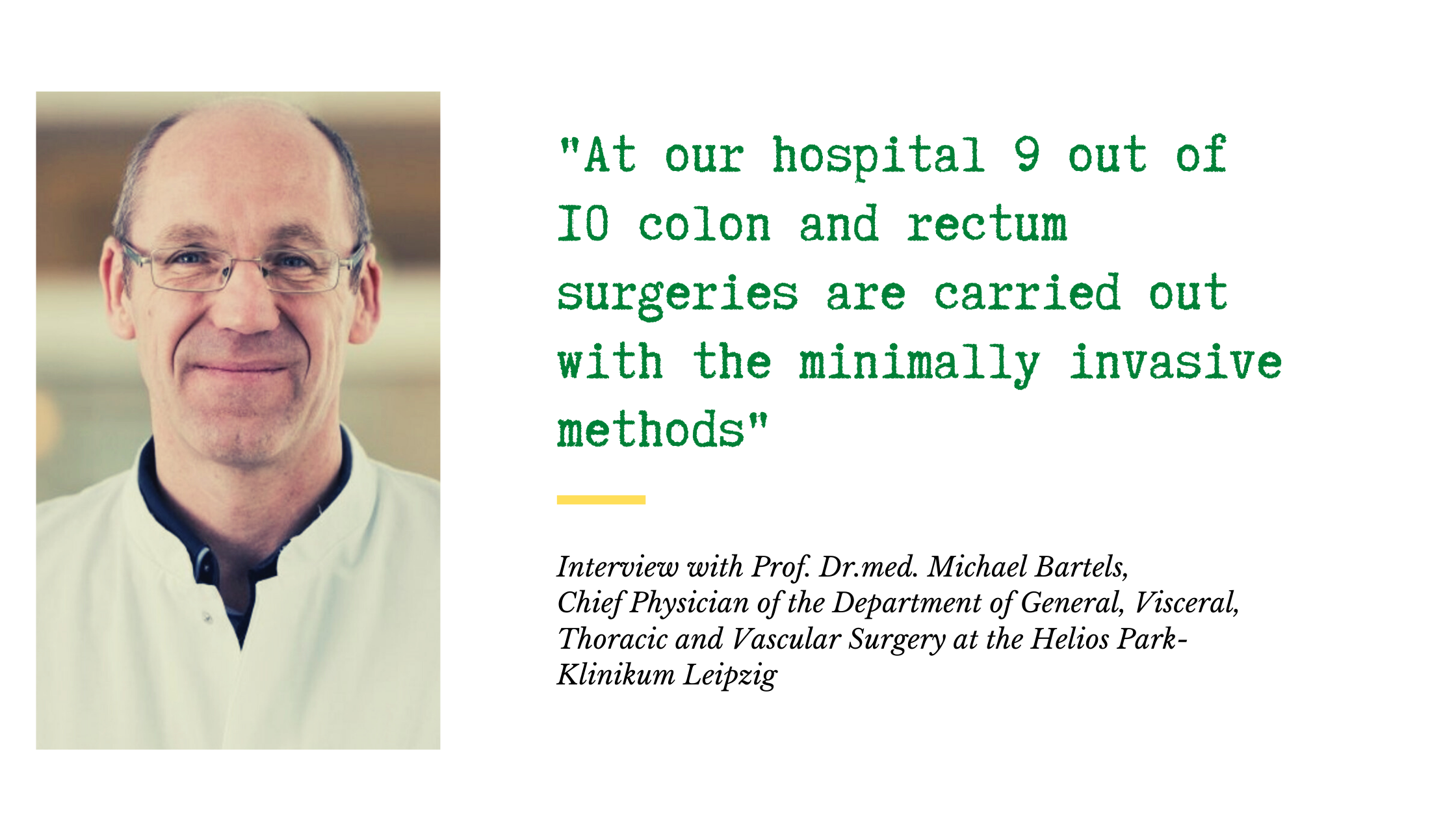
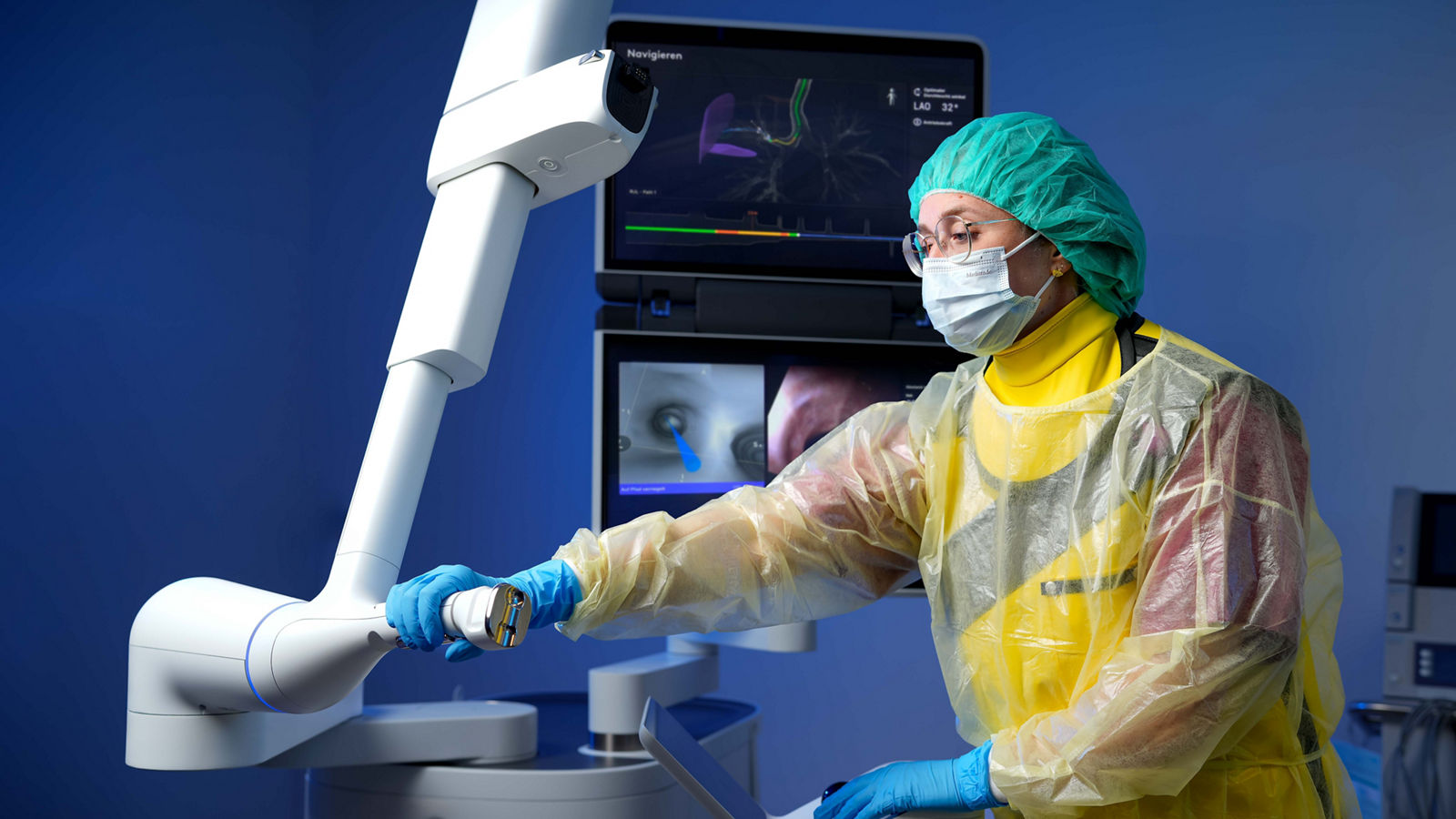
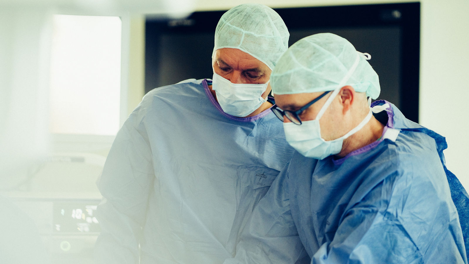
Esophagectomy is one of the most complex procedures in oncological surgery and requires considerable experience at a specialized center. Today, the operation can be performed using open, minimally invasive, or robot-assisted techniques.
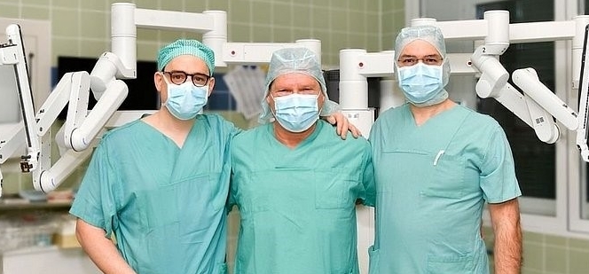
At Helios University Hospital Wuppertal, a DKG-certified Esophageal Cancer Center and the specialized Center West for Esophageal and Gastric Surgery provide the full spectrum of esophageal surgery, including minimally invasive and robot-assisted procedures. Before major surgery, patients may also undergo an individualized prehabilitation program designed to improve their physical condition before the procedure.
Helios Hospital Krefeld also specializes in surgery for esophageal cancer. Procedures are generally performed using minimally invasive techniques and predominantly with robotic assistance. The center cooperates with University Hospital Cologne: complex surgical cases can be discussed jointly, and specialists from Cologne may participate in procedures performed in Krefeld.
Chemotherapy before and after surgery
For many locally advanced tumors, surgery alone is not sufficient because microscopic cancer cells may already be present outside the primary tumor or in the lymph nodes.
For this reason, perioperative chemotherapy — systemic treatment before surgery and, depending on the clinical situation, after surgery — is used for certain forms of esophageal cancer. This approach is particularly important for adenocarcinomas of the lower esophagus and the gastroesophageal junction.
Preoperative treatment can reduce the size of the tumor, target possible micrometastases, and increase the likelihood of complete surgical removal. After surgery, the need for further systemic treatment is determined according to the original treatment strategy and the histological findings from the resected tumor.
Chemoradiotherapy
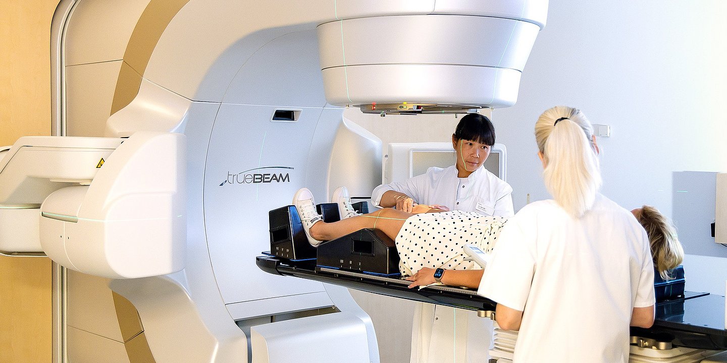
In chemoradiotherapy, radiation therapy is administered together with anticancer drugs. Chemotherapy not only acts systemically against cancer cells but can also make them more sensitive to radiation.
Chemoradiotherapy may be used before surgery for locally advanced esophageal cancer. This approach plays a particularly important role in the treatment of squamous cell carcinoma.
In some situations, chemoradiotherapy is used as the primary treatment without subsequent surgery. This is known as definitive chemoradiotherapy and may be appropriate for certain squamous cell carcinomas, particularly tumors located in the upper esophagus, as well as when surgery is not possible or would carry an excessively high risk.
At a specialized center, the choice between surgery following preoperative treatment and definitive chemoradiotherapy is made individually based on the tumor’s location, histological type and extent, as well as the patient’s overall condition.
Immunotherapy
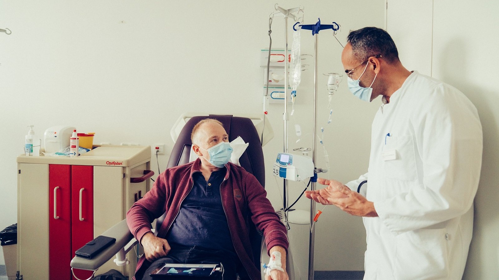
Immunotherapy has become an important part of modern treatment for advanced esophageal cancer. Immune checkpoint inhibitors help restore the ability of T cells to recognize and attack cancer cells.
These drugs are used primarily for locally advanced unresectable or metastatic disease, either alone or in combination with chemotherapy, depending on the type of tumor, the line of treatment, and its molecular and immunological characteristics.
PD-L1 expression may be taken into account when selecting treatment. Other tumor characteristics, including MSI/dMMR status, may also influence the choice of systemic therapy in certain patients.
Immunotherapy can also be used after preoperative chemoradiotherapy and surgery in selected patients when histological examination shows residual tumor. Its role is therefore no longer limited to metastatic disease.
At Helios Hospital Berlin-Buch, patients with esophageal tumors receive systemic treatment within a specialized center for gastrointestinal tumors. Treatment options include chemotherapy, immunotherapy, surgery, and endoscopic procedures.
Targeted therapy
In advanced esophageal cancer, systemic treatment increasingly depends on the biological characteristics of the individual tumor.
One of the most important examples is HER2. Overexpression of this protein or amplification of the corresponding gene occurs in a proportion of adenocarcinomas of the esophagus and gastroesophageal junction. In patients with HER2-positive tumors, drugs directed against this molecular target may be incorporated into systemic treatment.
For this reason, biomarker testing of tumor tissue is an important part of treatment planning in advanced disease. The results of HER2, PD-L1, and other molecular tests can help determine whether standard chemotherapy should be supplemented with targeted therapy or immunotherapy.
Rehabilitation after esophageal resection
Recovery after esophagectomy takes time because the operation changes the anatomy of the upper digestive tract and the normal process of eating. During the first days after surgery, the main priorities are pain management, respiratory therapy, and early mobilization. These measures help reduce the risk of postoperative complications and gradually restore physical activity.
Nutritional support is particularly important. Oral food intake is resumed gradually, while tube feeding may be used during the early recovery period if necessary. After surgery, patients are generally advised to eat small meals several times a day, chew food thoroughly, and ensure adequate protein and calorie intake. Nutritional counseling can help patients adapt their diet and prevent excessive weight loss.
During the first few months, patients may experience early satiety, reflux, reduced appetite, or dumping syndrome, which can cause weakness, sweating, palpitations, or digestive symptoms after meals. In many cases, these problems can be reduced by adjusting the diet and eating habits.
Another possible consequence of surgery is anastomotic narrowing at the site where the remaining esophagus is connected to the reconstructed digestive tract. If this causes difficulty swallowing, the narrowed area can often be widened endoscopically.
Physical activity is increased gradually according to the patient’s condition. Specialized oncological rehabilitation may include physiotherapy, breathing exercises, nutritional support, and measures to restore physical strength and endurance. Regular follow-up helps monitor body weight and nutritional status, identify postoperative problems at an early stage, and support the patient’s return to everyday life.
Esophageal cancer treatment at Helios Hospitals
Esophageal cancer treatment at Helios is based on a multidisciplinary and multimodal approach. Each case is reviewed by specialists from different disciplines, including gastroenterology, oncological surgery, medical oncology, radiation oncology, radiology, and pathology. This makes it possible to determine the optimal sequence of treatment — from endoscopic procedures for early-stage tumors to combinations of systemic therapy, radiation therapy, and surgery for locally advanced disease.
One of the key advantages of Helios is the extensive use of minimally invasive techniques. In selected patients, early tumors can be removed endoscopically using EMR or ESD, avoiding major surgery. When esophageal resection is required, specialized centers offer minimally invasive and robot-assisted procedures, enabling complex surgical steps to be performed with a high degree of precision.
Particular attention is paid to personalized systemic treatment. In advanced disease, tumor tissue can be tested for biomarkers that may influence treatment selection. Depending on the tumor type and molecular findings, treatment may include immunotherapy and targeted therapies in addition to chemotherapy.
Cancer treatment at Helios extends beyond controlling or removing the tumor. Nutritional support, preparation for major surgery, postoperative recovery, and oncological rehabilitation are important components of comprehensive care. This is particularly relevant for patients with esophageal cancer, as both the disease and its treatment can significantly affect swallowing, nutrition, body weight, and physical strength.
The key advantage of treatment at Helios is the ability to combine advanced endoscopy, high-tech surgery, radiation and systemic therapy, molecular diagnostics, and rehabilitation within an individualized treatment plan.
If you are interested in arranging diagnosis or treatment at one of the Helios hospitals, you can submit a request using the contact form on our website or contact us via the email address provided at the top of the website.
Learn more about arranging treatment at Helios hospitals ⟶
Do you need more information about Helios Hospitals or want to schedule your treatment?