Pancreatic cancer treatment: Modern methods and treatment options
- Early diagnosis of the disease
- Causes and risk factors
- Symptoms of pancreatic cancer
- Diagnosis of pancreatic cancer
- Pancreatic cancer treatment
- Individualized treatment and advanced options at Helios
The pancreas is one of the largest glands in the human body. It produces essential hormones that regulate blood sugar levels (endocrine function), as well as digestive secretions containing various enzymes that help break down food (exocrine function). Anatomically, the pancreas is located in the retroperitoneal space, behind the peritoneum and in front of the major blood vessels (the aorta and inferior vena cava). It is divided into four regions: the head, neck, body, and tail.
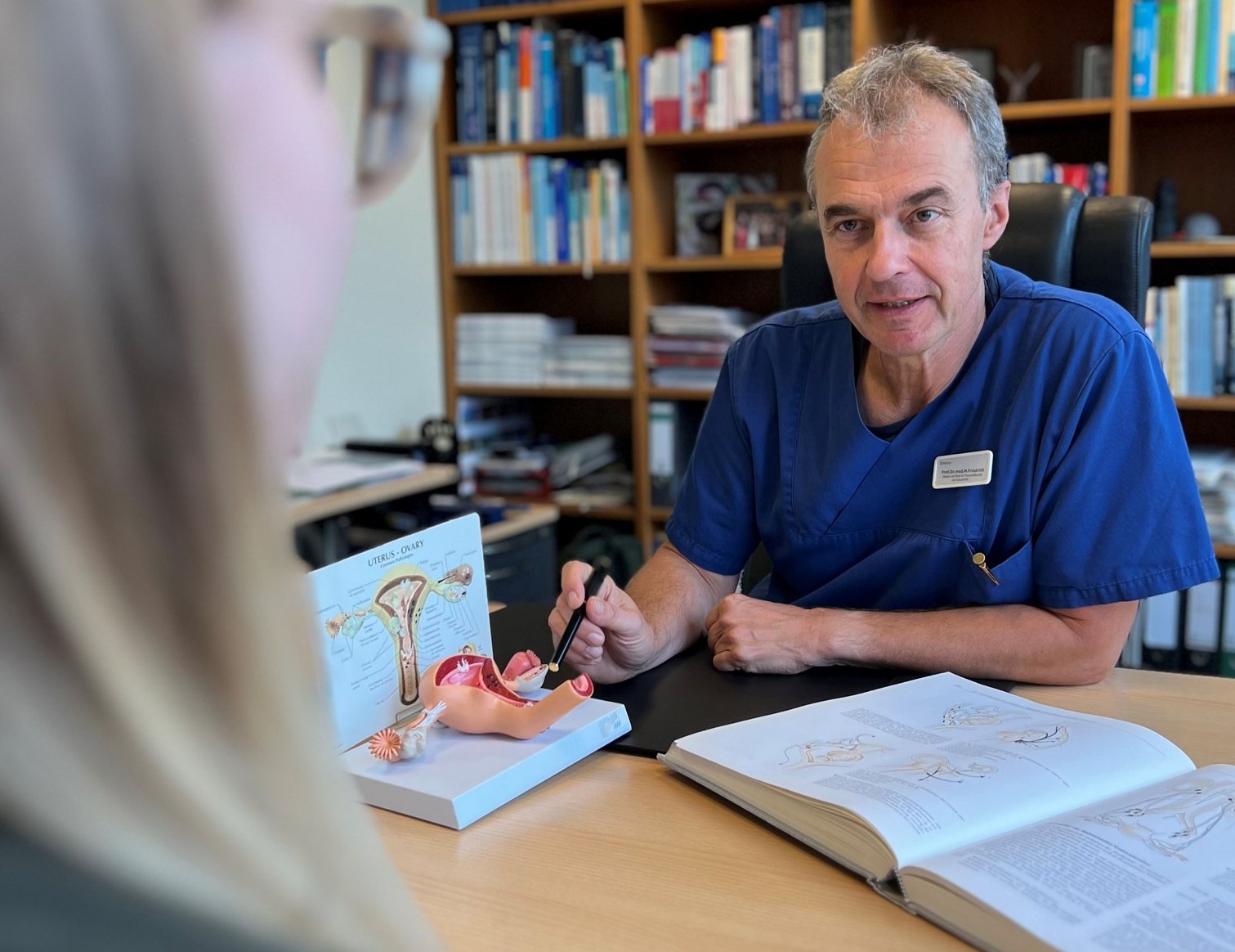
“Pancreatic cancer is difficult to treat because it is often diagnosed at an advanced stage and may spread unnoticed before it is detected. The disease occurs predominantly in older adults. The average age at diagnosis is 76 years for women and 72 years for men. In both men and women, pancreatic cancer is the fourth leading cause of cancer-related death, in Germany” – Dr. Jale Celebi, senior physician and specialist in visceral surgery and coordinator of the Pancreatic Center at Helios Hospital Berlin-Buch.
The common bile duct passes behind the head of the pancreas and enters the duodenum together with the main pancreatic duct. The openings of these two ducts are protected against the backflow of food and gastric contents by a circular sphincter (the sphincter of Oddi / ampulla of Vater). The duodenum surrounds the head of the pancreas in a C-shape and is closely attached to it.
The wall of this segment of the intestine receives its blood supply from arteries associated with the head of the pancreas. Because of the close anatomical relationship between the pancreas, particularly the duodenum and the common bile duct, pancreatic cancer treatment may require partial or complete removal of these structures. However, accurately distinguishing between benign and malignant tumors is essential for precise surgical planning. In addition to monitoring typical tumor markers associated with pancreatic or bile duct cancer, specific markers may be used to identify or rule out tumors arising from the hormone-producing cells of the pancreas. These are known as neuroendocrine tumors (NETs) and may originate, for example, from insulin-producing cells.
Early diagnosis of the disease
For people without symptoms and without a significantly increased risk of pancreatic cancer, routine screening is currently not recommended. This also applies to people with conditions that only moderately increase the risk, such as diabetes mellitus, gallstones, or fatty liver disease with inflammation.
People with a significantly increased risk of developing pancreatic cancer may be offered regular surveillance examinations. This group includes, among others, people:
- with several first-degree relatives who have been diagnosed with pancreatic cancer;
- who carry certain genetic mutations;
- with rare hereditary conditions such as Peutz–Jeghers syndrome;
- or with hereditary chronic pancreatitis.
The main methods used for surveillance of high-risk individuals are magnetic resonance imaging (MRI) and endoscopic ultrasound (EUS). The age at which regular surveillance should begin depends on the individual risk. It often starts between the ages of 40 and 55, or ten years before the age at which the youngest affected family member was diagnosed with pancreatic cancer.
Causes and risk factors
The exact causes of pancreatic cancer are not yet fully understood. The disease develops as a result of changes in pancreatic cells that cause them to grow uncontrollably and form a tumor. However, a number of factors are known to increase the risk of developing pancreatic cancer.
The main risk factors include:
- Age. The risk of pancreatic cancer increases with age. Most patients are diagnosed after the age of 65.
- Smoking. Tobacco use is one of the most significant preventable risk factors. People who smoke are approximately twice as likely to develop pancreatic cancer as those who have never smoked.
- Alcohol. Regular heavy alcohol consumption may increase the risk of pancreatic cancer. Excessive alcohol consumption is also one of the causes of chronic pancreatitis, which is itself an established risk factor for pancreatic cancer.
- Overweight and obesity. Obesity is associated with an increased risk of pancreatic cancer, partly due to metabolic changes in the body.
- Diabetes. Pancreatic cancer is more common in people with diabetes, particularly type 2 diabetes. At the same time, newly diagnosed diabetes can sometimes be not only a risk factor but also an early sign of pancreatic cancer.
- Chronic pancreatitis. Long-term inflammation of the pancreas increases the risk of developing pancreatic cancer. The risk may be particularly high in people with certain hereditary forms of pancreatitis.
- Family history and genetic factors. The risk is higher in people who have close relatives with pancreatic cancer. Inherited variants in genes such as BRCA1, BRCA2, PALB2, ATM, and others may also increase the risk, as can hereditary conditions such as Lynch syndrome and Peutz–Jeghers syndrome.
Having one or even several risk factors does not mean that a person will necessarily develop pancreatic cancer. Conversely, the disease can occur in people with no known risk factors. However, individuals with a strong family history of pancreatic cancer or certain inherited genetic variants may be advised to undergo genetic counseling and regular surveillance.
Symptoms of pancreatic cancer
“Unfortunately, pancreatic tumors often do not cause symptoms until the disease has reached an advanced stage,” explains Dr. Jale Celebi. “Some patients, for example, seek medical attention because of nonspecific pain in the upper abdomen or back. Further examination may then reveal that pancreatic cancer is the underlying cause of these symptoms.”
One possible sign of the disease is new-onset diabetes, particularly when it develops without an obvious cause. The pancreas produces insulin and plays an important role in regulating blood sugar levels, so a tumor can interfere with this function. However, developing diabetes does not in itself mean that a person has pancreatic cancer.
Another characteristic symptom is jaundice, particularly when the tumor is located in the head of the pancreas. The common bile duct, which carries bile into the duodenum, runs alongside this part of the pancreas. As the tumor grows, it can compress or block the bile duct, preventing bile from draining normally and causing bilirubin levels in the blood to rise. This may initially cause yellowing of the whites of the eyes, followed by yellowing of the skin. This type of jaundice is often painless and may be one of the first noticeable signs of pancreatic cancer.
Pancreatic cancer may also cause changes in the color of urine and stool: urine may become dark, while stools may become pale or clay-colored.
At more advanced stages, other symptoms and signs may include:
- nonspecific pain and digestive problems;
- fluid accumulation in the abdomen (ascites);
- inflammation of the pancreas (pancreatitis);
- blood clots and thrombosis.
Diagnosis of pancreatic cancer
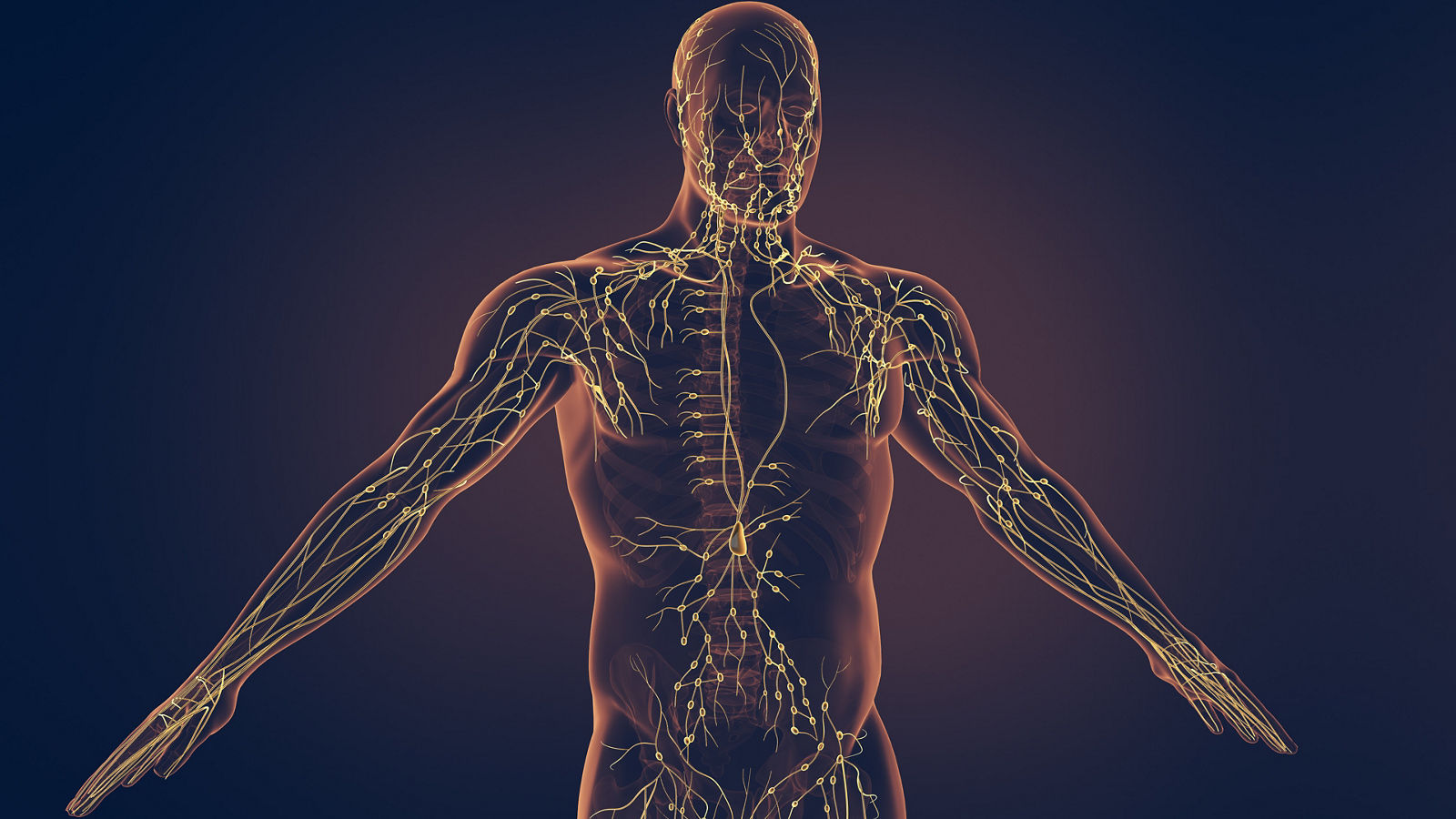
When pancreatic cancer is suspected, diagnostic testing is aimed not only at detecting the tumor. It is also important to determine its exact location and size, assess the surrounding tissues, lymph nodes, and blood vessels, identify possible metastases, and establish whether the tumor can be surgically removed.
“One of the most important factors in the treatment of a pancreatic tumor is diagnosing the disease as early as possible. The smaller the tumor, the better the chances of successfully treating the cancer.” — Prof. Dr. Marc H. Jansen, Head of the Department of General, Visceral and Minimally Invasive Surgery and Director of the Visceral Oncology Center at Helios Hospital Emil von Behring.
The diagnostic process usually begins with a consultation and physical examination. The doctor will ask about the nature and duration of symptoms, underlying medical conditions, and possible risk factors. Blood tests are also performed, including tests to assess liver and biliary function. Tumor markers, particularly CA 19-9, may also be measured. However, tumor markers alone cannot confirm a diagnosis of pancreatic cancer and must always be interpreted together with the results of other examinations.
Various imaging techniques are used to diagnose pancreatic cancer:
- Ultrasound is often one of the first imaging examinations performed, particularly in patients with abdominal pain or signs of impaired bile flow. However, because of the location of the pancreas, conventional ultrasound does not always provide a sufficiently detailed view of the organ.
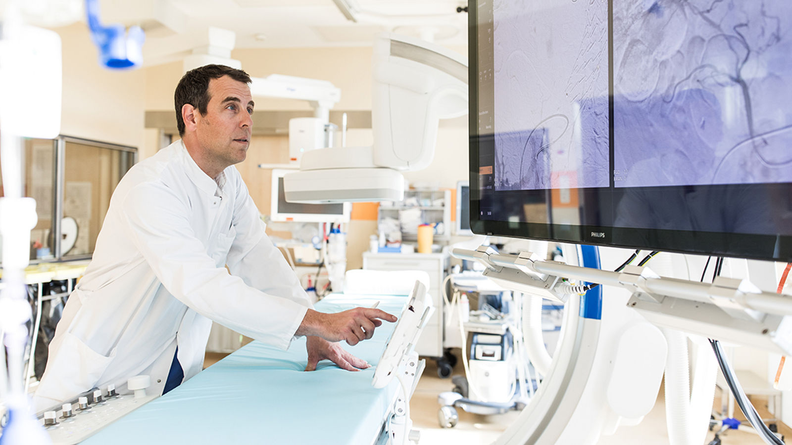
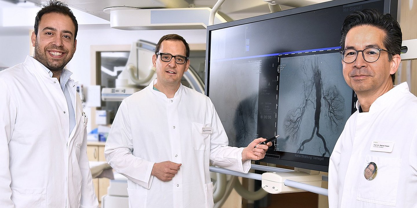
- Computed tomography (CT) can determine the location and size of the tumor, assess its relationship with major blood vessels and neighboring organs, and detect possible metastases. This information is particularly important when planning surgery.
- Magnetic resonance imaging (MRI) provides detailed images of the pancreas and surrounding structures. A specialized MRI technique known as magnetic resonance cholangiopancreatography (MRCP) allows detailed visualization of the pancreatic and bile ducts.
- Endoscopic ultrasound (EUS) allows the pancreas to be examined at close range through the wall of the stomach and duodenum. If necessary, a targeted biopsy can be performed during the procedure to obtain a tissue sample for histopathological examination.
- Endoscopic retrograde cholangiopancreatography (ERCP) is used to examine the bile and pancreatic ducts. Today, this procedure is particularly important when an obstruction of bile flow needs not only to be identified but also treated, for example by placing a stent.
A biopsy is not required in every case. If imaging findings clearly indicate a resectable tumor, surgery may in some cases be performed without a prior biopsy. Obtaining a tissue sample is particularly important when imaging findings are inconclusive or before systemic anticancer therapy is initiated.
Determining the stage of pancreatic cancer
Once the diagnosis has been confirmed, the extent of the disease is assessed through a process known as staging. The international TNM classification is used for this purpose. It takes into account the characteristics of the primary tumor (T), involvement of regional lymph nodes (N), and the presence of distant metastases (M). Based on these findings, pancreatic cancer is classified into stages ranging from I to IV.
An especially important factor in treatment planning is whether the tumor is resectable, meaning that it can be completely removed surgically. In the absence of distant metastases, a tumor may be classified as resectable, borderline resectable, or locally advanced and unresectable. Whether surgery is possible depends not only on the size of the tumor, but particularly on its location and its involvement with major blood vessels.
The results of these examinations help determine the stage of the disease and guide the choice of treatment. At specialized Helios cancer centers, cases of pancreatic cancer are reviewed by a multidisciplinary team that includes surgeons, gastroenterologists, oncologists, radiologists, and other specialists.
Pancreatic cancer treatment
The choice of treatment for pancreatic cancer depends primarily on the stage of the disease and whether the tumor can be completely removed surgically. Depending on the individual clinical situation, treatment may include surgery, chemotherapy, radiation therapy, or a combination of several approaches. For localized and resectable tumors, surgery is the main treatment option, while borderline resectable, locally advanced, or metastatic pancreatic cancer may require different systemic and local treatment strategies.
Surgical treatment
If the tumor is localized and can be completely removed, surgery is the primary treatment option that may offer the possibility of long-term disease control and potentially a cure. The type of surgery depends mainly on the location of the tumor.
For tumors located in the head of the pancreas, a pancreaticoduodenectomy, also known as the Whipple procedure, is usually performed. This involves removing the head of the pancreas together with the duodenum, part of the bile duct, the gallbladder, and regional lymph nodes. Depending on the surgical technique, part of the stomach may be preserved. After the tumor has been removed, the surgeon reconstructs the digestive tract to restore the flow of bile and pancreatic secretions.
For tumors located in the body or tail of the pancreas, a distal pancreatectomy is usually performed, typically together with removal of the spleen and regional lymph nodes.
In selected cases, a total pancreatectomy, meaning complete removal of the pancreas, may be necessary. Following this procedure, patients require lifelong insulin therapy and pancreatic enzyme replacement.
Extended surgery with vascular reconstruction
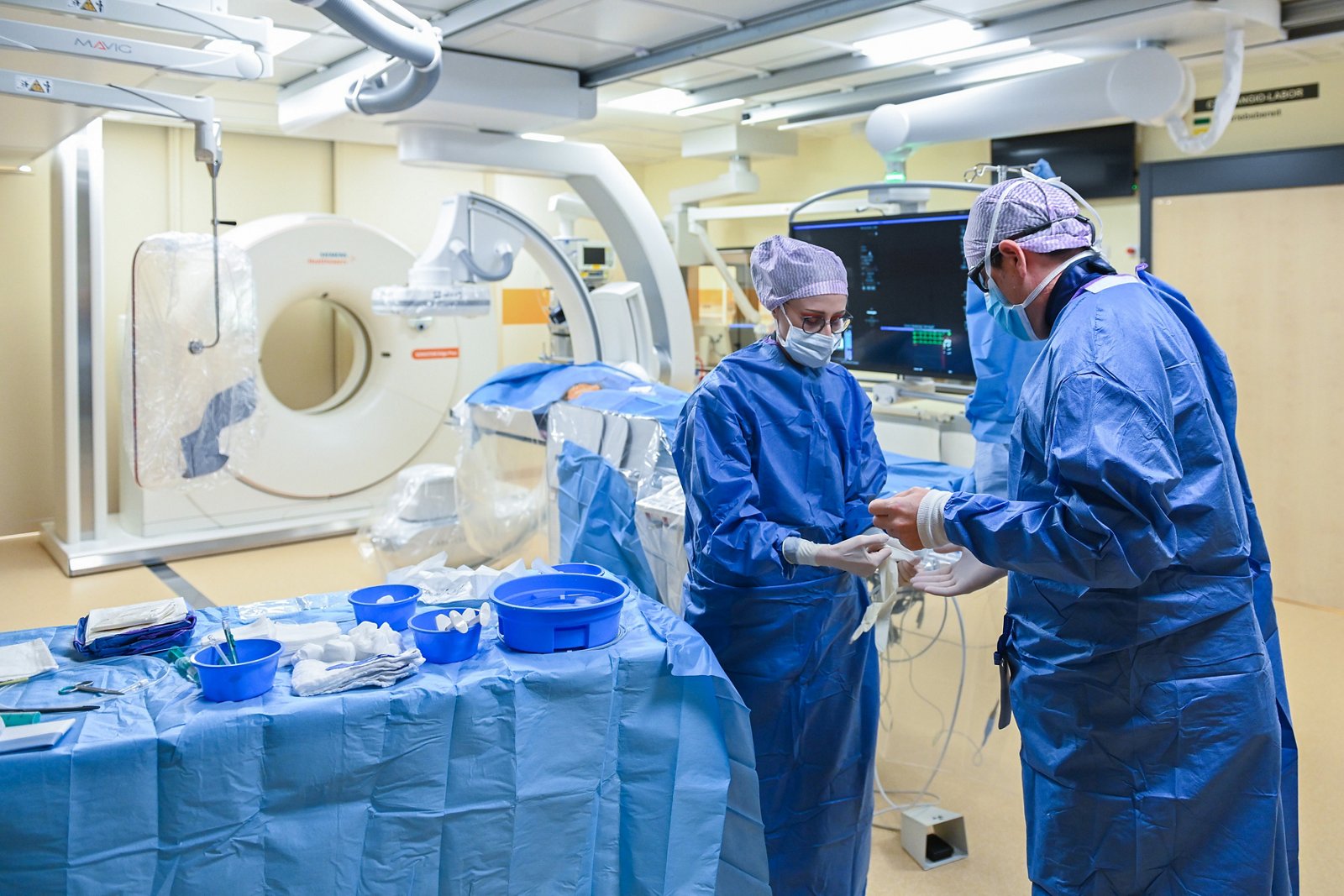
When a tumor involves major blood vessels, surgical treatment becomes considerably more complex and requires extensive expertise in both pancreatic and vascular surgery. For this reason, such procedures are primarily performed at specialized centers.
At Helios HSK Wiesbaden, selected patients with vascular involvement may undergo extended surgery in which the affected segment of the portal vein or superior mesenteric vein is removed together with the tumor and the blood vessel is subsequently reconstructed. This approach may make radical surgical treatment an option in certain complex cases where standard tumor resection would not be technically feasible.
The decision to perform surgery with vascular reconstruction is made on an individual basis and depends on the extent of the tumor, the type and degree of vascular involvement, and whether complete removal of the tumor can be achieved.
Robot-assisted surgery
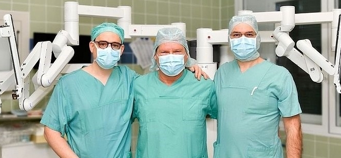
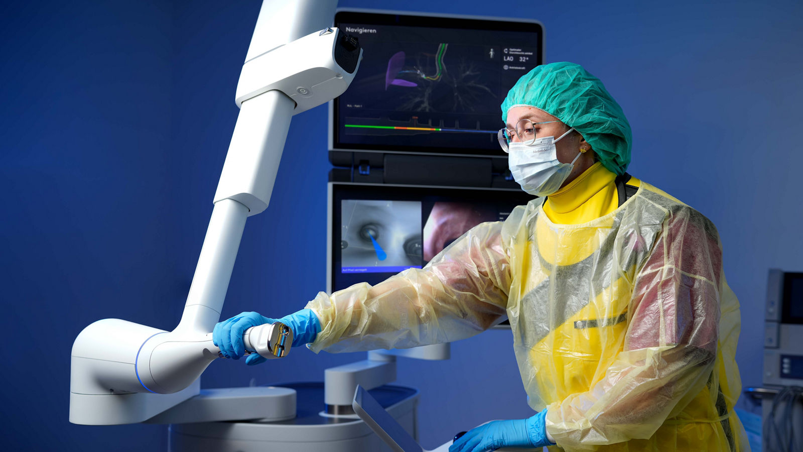
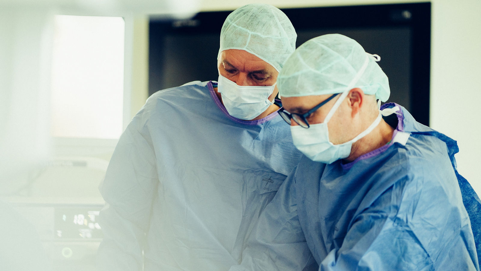
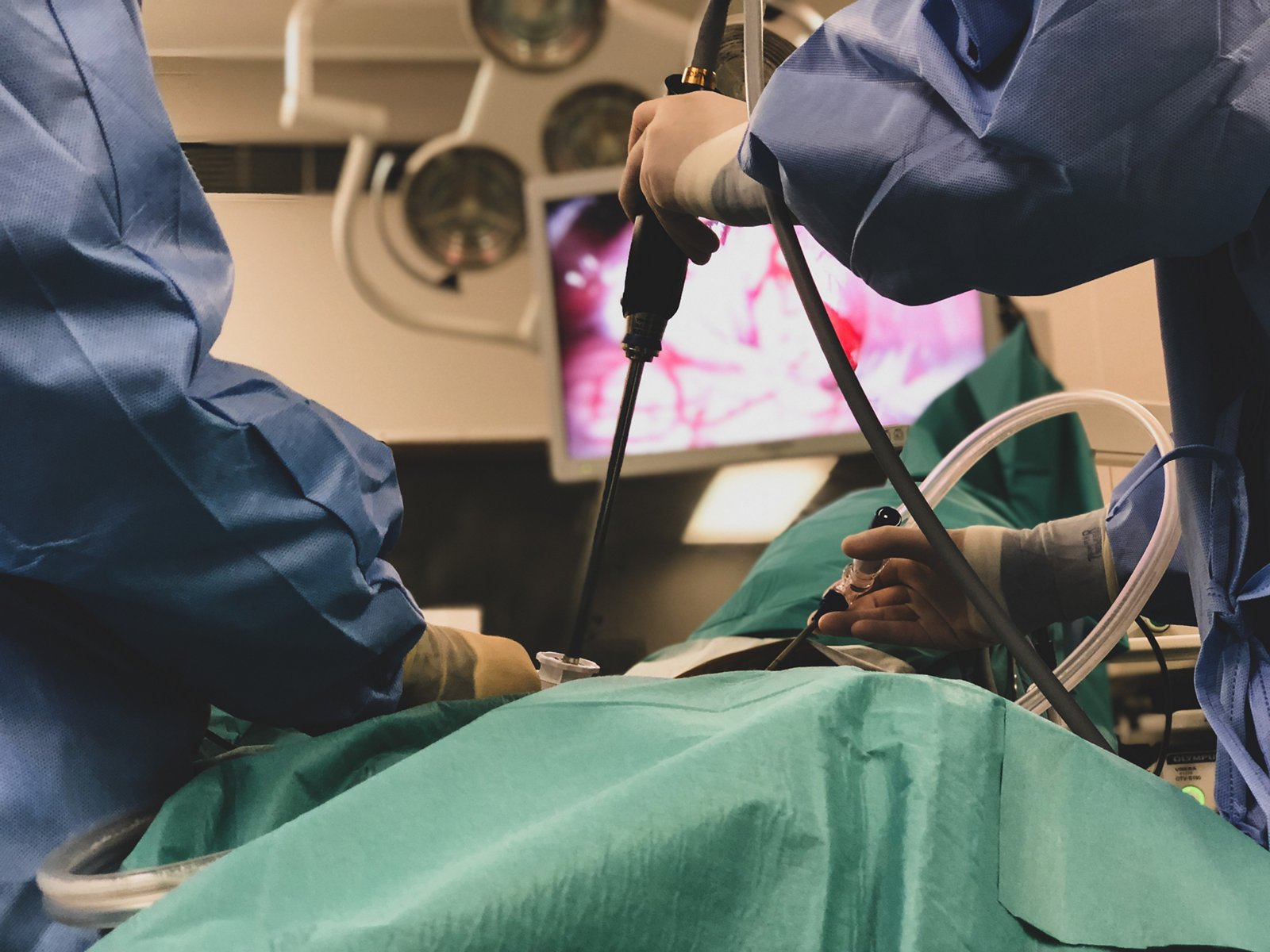
Some pancreatic procedures can be performed using minimally invasive techniques, either laparoscopically or with the Da Vinci robotic surgical system. During robot-assisted surgery, the surgeon controls the instruments from a console and operates with a magnified three-dimensional view of the surgical field. The high precision and extensive range of motion of the robotic instruments enable complex surgical procedures to be performed through small incisions.
Robot-assisted pancreatic surgery is available at several Helios hospitals, including Helios HSK Wiesbaden, Helios Hospital Krefeld, Helios Park Hospital Leipzig, Helios Hospital Berlin-Buch, and Helios Hospital Emil von Behring.
Compared with open surgery, robot-assisted procedures are performed through smaller incisions and may be associated with less blood loss, reduced postoperative pain, and faster recovery. Enhanced visualization of the surgical field and the high degree of mobility of the robotic instruments help the surgeon operate precisely around blood vessels and other important anatomical structures. Whether robot-assisted surgery is appropriate is determined individually based on the location and extent of the tumor and the complexity of the required procedure.
Chemotherapy
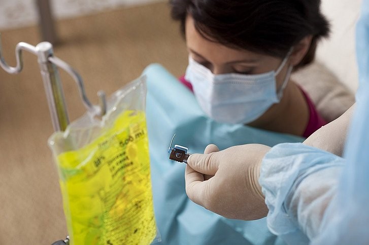
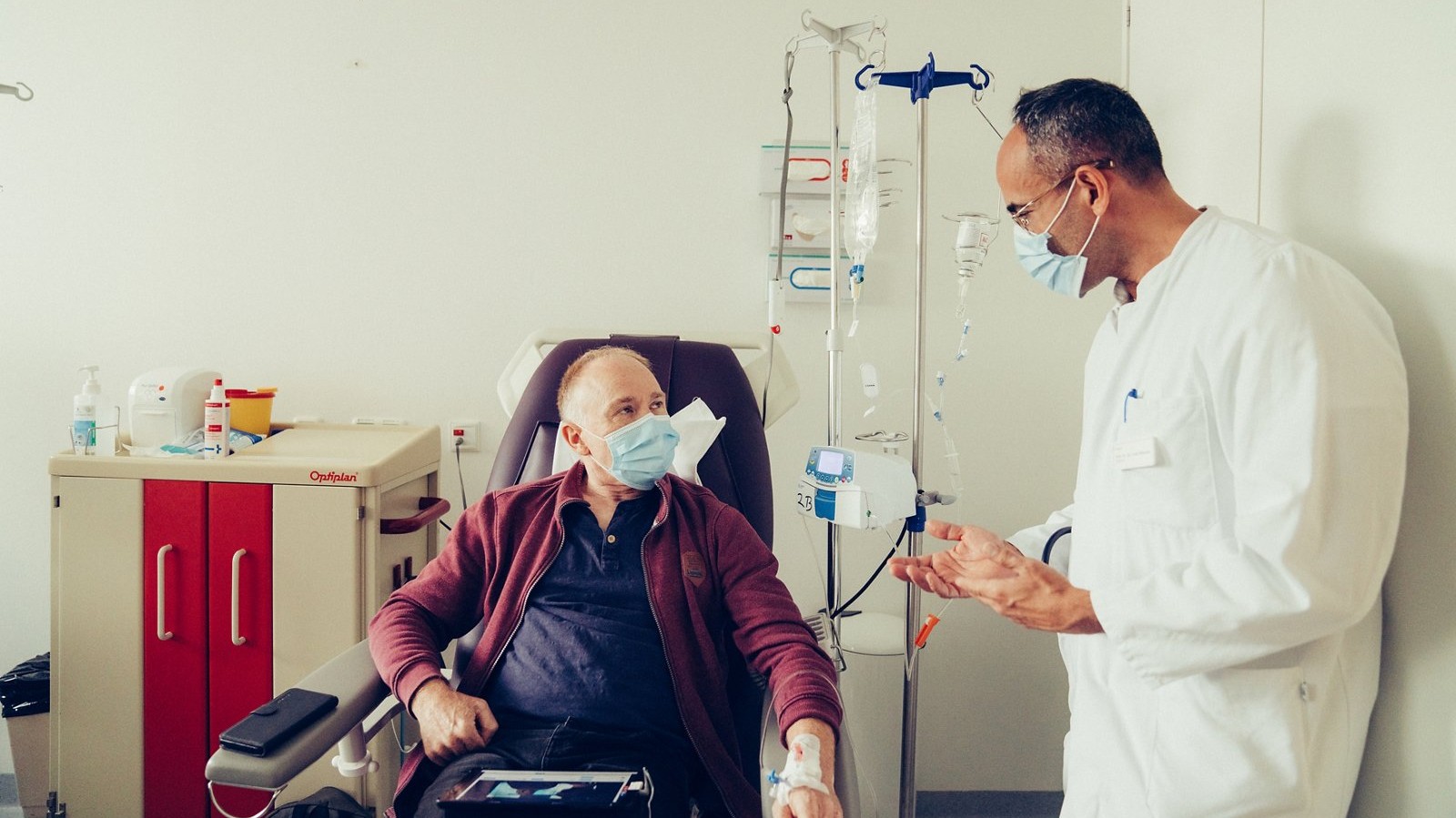
Chemotherapy is one of the main treatments for pancreatic cancer and may be used on its own or in combination with surgery or radiation therapy. It involves anticancer drugs that target cancer cells throughout the body. The drugs are usually administered intravenously in treatment cycles, with scheduled breaks that allow the body to recover between cycles.
The choice of chemotherapy regimen depends on the stage of the disease, the patient’s overall health, previous treatment, and other individual factors. Depending on the clinical situation, chemotherapy may be given before surgery (neoadjuvant therapy), after surgery (adjuvant therapy), or used as the primary treatment for advanced disease.
Chemotherapy before surgery
Neoadjuvant chemotherapy is given before surgery. It is particularly important for borderline resectable tumors that are in contact with major blood vessels or partially involve them.
The aim of this treatment is to target the tumor and possible microscopic cancer deposits before surgery, assess how the tumor responds to therapy, and increase the likelihood of complete surgical removal. In some cases, treatment may reduce the extent of the disease sufficiently to make surgery possible when it was initially considered technically difficult or not feasible.
After several treatment cycles, imaging and other examinations are repeated. The results help assess the tumor’s response to treatment and determine whether surgery can be performed.
Chemotherapy after surgery
Adjuvant chemotherapy is given after surgical removal of the tumor. Even when surgery is successful and all visible tumor tissue has been removed, individual cancer cells or microscopic deposits that cannot be detected with current imaging techniques may remain in the body.
The main goal of adjuvant chemotherapy is to destroy any remaining cancer cells and thereby reduce the risk of the cancer returning. The treatment regimen and duration are selected individually based on the histopathology results, the patient’s overall condition, and recovery after surgery.
Irreversible electroporation (IRE / NanoKnife)
For selected patients with locally advanced, unresectable pancreatic cancer, irreversible electroporation (IRE), also known as NanoKnife, may be considered. This is a local tumor treatment in which special electrodes deliver short, high-voltage electrical pulses that disrupt the membranes of cancer cells, ultimately causing them to die.
Unlike thermal ablation techniques, IRE does not rely on heating the tumor tissue. This makes it possible to treat tumors located in close proximity to major blood vessels and other important anatomical structures.
IRE treatment for pancreatic tumors is available, among others, at Helios Hanseatic Hospital Stralsund and Helios Hospital Krefeld. The Helios hospital in Stralsund has extensive experience with IRE for unresectable pancreatic cancer and has been using this technology since 2015.
IRE does not replace standard treatments for pancreatic cancer and is not suitable for every patient. Whether it is an appropriate option is determined individually after assessing the extent of the disease, the location of the tumor, vascular involvement, and the results of previous treatment.
Nuclear medicine
Nuclear medicine treatments may be used for certain pancreatic neuroendocrine tumors (pNETs). These tumors arise from neuroendocrine cells and differ from the most common type of pancreatic cancer — pancreatic ductal adenocarcinoma — in their biological characteristics and treatment approaches.
One specialized treatment is peptide receptor radionuclide therapy (PRRT). During this treatment, the patient receives a radiopharmaceutical containing a radioactive isotope, such as lutetium-177, linked to a somatostatin analogue. The drug binds to somatostatin receptors on the surface of tumor cells and delivers radiation directly to the tumor and its metastases, helping to limit exposure to surrounding healthy tissues.
PRRT is used only for tumors that express sufficient levels of the relevant receptors. Before treatment, receptor expression is assessed using specialized molecular imaging, such as PET/CT with radiolabeled somatostatin analogues.
Radionuclide therapy for neuroendocrine tumors is available, among other locations, at the Department of Nuclear Medicine at Helios Hospital Berlin-Buch. Whether PRRT is an appropriate treatment option is determined individually based on the type and extent of the tumor, somatostatin receptor expression, and previous treatments.
Radiation therapy
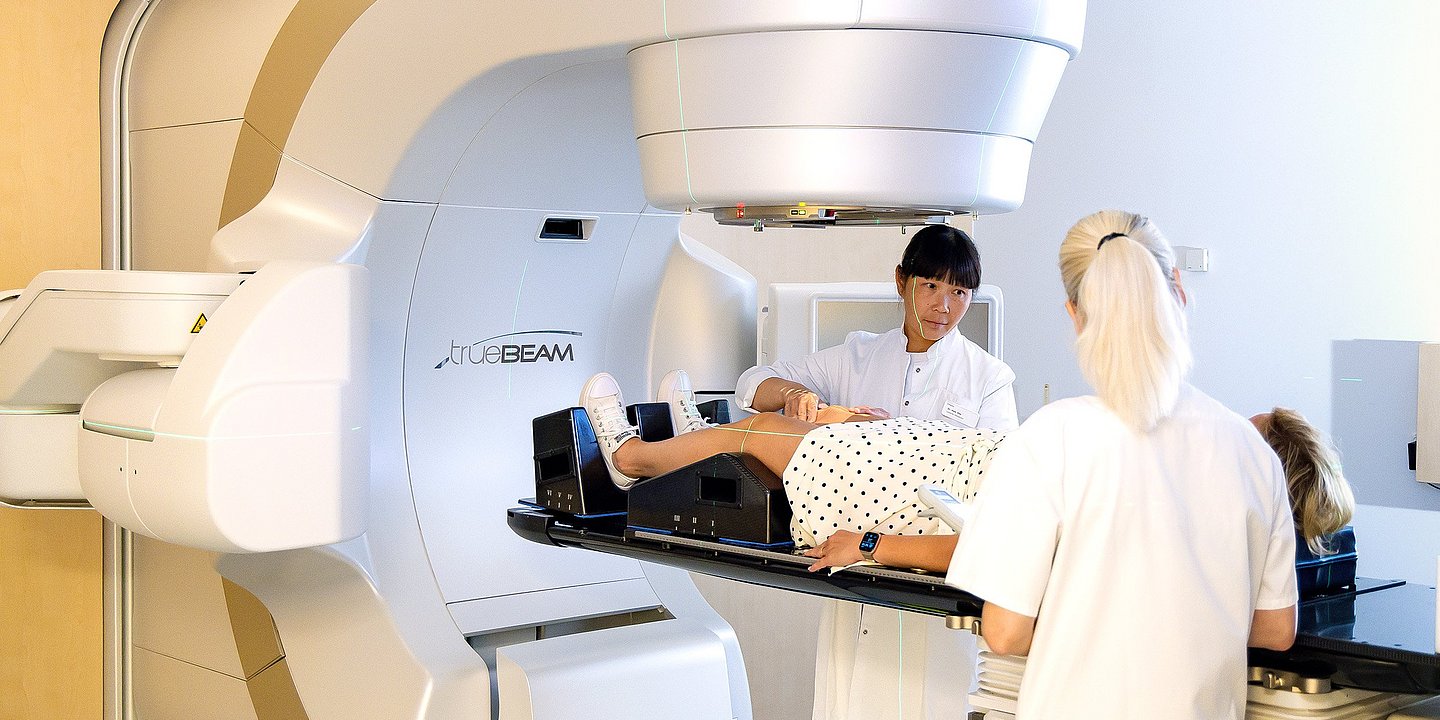
Radiation therapy is not a primary treatment for pancreatic cancer, but it may be used in selected patients, usually in combination with chemotherapy.
For locally advanced pancreatic cancer without distant metastases, radiation therapy may be used to achieve local tumor control, including after a course of systemic therapy. In some cases, radiation therapy may also be considered as part of a multimodal treatment strategy for borderline resectable tumors.
In advanced disease, radiation therapy may be used for palliative purposes, for example to relieve pain and other symptoms caused by the tumor or metastases. The decision to use radiation therapy is made individually, taking into account the stage of the disease, the location of the tumor, and previous treatment.
Follow-up care and rehabilitation
After completing treatment for pancreatic cancer, patients require regular medical follow-up. The aim is to monitor recovery, detect possible recurrence or metastases at an early stage, and manage any long-term effects of treatment.
The follow-up schedule is tailored to the individual patient based on the stage of the disease, the treatment received, and their overall health. Follow-up may include appointments with an oncologist and other specialists, laboratory tests, monitoring of the tumor marker CA 19-9, and imaging examinations, primarily CT scans, with MRI or other diagnostic tests performed when necessary. The frequency of these examinations may change over time and is determined by the treating medical team.
Nutritional support is an important part of recovery after pancreatic surgery. Removal of part or all of the pancreas can reduce the production of digestive enzymes, which may lead to problems digesting food, weight loss, and nutritional deficiencies. In such cases, pancreatic enzyme replacement therapy may be prescribed, together with an individually tailored nutritional plan.
Blood glucose levels also need to be monitored after surgery, as a reduction in functioning pancreatic tissue can lead to the development or worsening of diabetes. After complete removal of the pancreas, patients require lifelong insulin therapy and pancreatic enzyme replacement.
Rehabilitation may include physical therapy and a gradual return to physical activity, nutritional counseling, management of pain and other symptoms, and psychological support. The goal is to help patients recover from intensive treatment, improve their nutritional and physical condition, and gradually return to everyday life.
Individualized treatment and advanced options at Helios
Pancreatic cancer treatment often requires a combination of different therapies and the expertise of specialists from several medical disciplines. At Helios pancreatic and cancer centers, complex cases are reviewed by multidisciplinary tumor boards to determine the most appropriate sequence of surgical and systemic treatment.
Depending on the individual clinical situation and the Helios center, patients may have access to complex pancreatic surgery with vascular reconstruction, minimally invasive and robot-assisted procedures, modern systemic therapies, radiation therapy, and specialized local treatments such as IRE. At selected Helios hospitals, patients may also have the opportunity to participate in clinical trials investigating new treatment approaches.
If you are interested in arranging diagnosis or treatment at one of the Helios hospitals, you can submit a request using the contact form on our website or contact us via the email address provided at the top of the website.
Learn more about arranging treatment at Helios hospitals ⟶
Read more:
Do you need more information about Helios Hospitals or want to schedule your treatment?