
Mitral valve replacement and repair: modern techniques
- When is mitral valve replacement or repair necessary
- Mitral valve repair techniques
- Mitral valve replacement techniques
- Percutaneous balloon mitral commissurotomy
- Life after mitral valve repair or replacement
- Mitral valve treatment at Helios hospitals
Mitral valve disease is one of the most common forms of valvular heart disease. According to estimates from international studies, mitral regurgitation affects approximately 2% of the population, with its prevalence increasing significantly with age. Thousands of mitral valve repair and replacement procedures are performed across Europe every year, while advances in minimally invasive and transcatheter technologies continue to expand treatment options for patients with different forms of the disease.
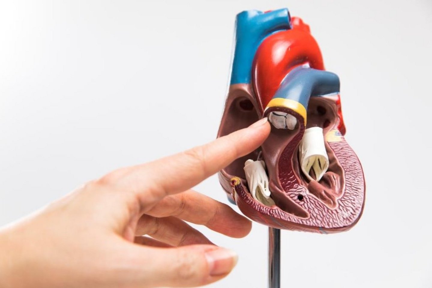
The mitral valve is located between the left atrium and the left ventricle and ensures that blood flows in one direction. When its leaflets do not close properly, some blood leaks back into the atrium, causing mitral regurgitation. When the valve opening becomes narrowed, mitral stenosis develops, restricting blood flow into the left ventricle. Both conditions can place additional strain on the heart, cause shortness of breath, fatigue, and heart rhythm disturbances, and gradually reduce quality of life.
Timely, high-quality mitral valve intervention is essential for preserving heart function. Modern cardiac surgery aims not only to restore normal blood flow but also, whenever possible, to preserve the patient's native valve. When the anatomy is suitable, mitral valve repair can restore the function of the valve leaflets and supporting structures without the need for a prosthesis. If repair is not feasible or is unlikely to provide a durable result, the valve can be replaced with a mechanical or biological prosthesis.
Selecting the most appropriate treatment requires accurate diagnosis and the expertise of a specialized medical team. In addition to conventional surgery, minimally invasive procedures performed through small incisions and transcatheter interventions such as MitraClip (TEER) are now available, allowing mitral regurgitation to be reduced without open-heart surgery. The choice between valve repair, replacement, and catheter-based treatment depends on the underlying mechanism of valve dysfunction, anatomical characteristics, heart muscle function, and coexisting medical conditions.
At Helios Hospitals in Germany, mitral valve disease is treated through a multidisciplinary approach involving cardiologists, cardiac surgeons, and cardiac imaging specialists. Careful procedural planning, individualized treatment selection, and follow-up care all play an important role in achieving successful outcomes.
To understand when mitral valve repair or replacement may be necessary, let's explore the main causes of mitral valve disease and the indications for surgical treatment.
When is mitral valve replacement or repair necessary?
Mitral valve replacement or repair may be necessary when a disease disrupts normal blood flow between the left atrium and the left ventricle. In a healthy heart, the mitral valve opens to allow blood to enter the ventricle and closes tightly when the ventricle contracts, preventing blood from flowing backward. If the valve leaflets become damaged, deformed, or lose their mobility, the heart has to work harder.
The main indications for intervention are severe mitral regurgitation and severe mitral stenosis. However, the need for surgery depends not only on the severity of valve damage but also on the presence of symptoms, the condition of the heart muscle, changes in the size of the heart chambers, and the risk of further deterioration in cardiac function.
Mitral valve insufficiency (mitral regurgitation)
Mitral regurgitation is a condition in which the valve leaflets do not close completely, allowing some blood to flow back into the left atrium when the left ventricle contracts. This increases the volume of blood the heart must handle and, over time, may lead to enlargement of the heart chambers, shortness of breath, reduced exercise tolerance, and heart rhythm disturbances.
There are two main forms of the disease:
- Primary (organic) mitral regurgitation results from direct damage to the valve leaflets, chordae tendineae, or other structures of the valve apparatus. Possible causes include degenerative changes, mitral valve prolapse, ruptured chordae, rheumatic valve disease, and infective endocarditis.
- Secondary (functional) mitral regurgitation develops as a result of changes in the geometry and function of the left ventricle or atrium. The valve itself may have no significant structural damage, but its leaflets no longer close properly because the heart chambers have enlarged or the supporting structures have shifted.
In patients with severe primary mitral regurgitation and suitable valve anatomy, mitral valve repair is often preferred because it preserves the patient's native valve tissue. In secondary mitral regurgitation, treatment of the underlying heart disease is optimized first. If significant regurgitation persists, surgical or transcatheter treatment options may be considered.
Mitral valve prolapse
Mitral valve prolapse is a condition in which one or both valve leaflets bulge backward into the left atrium when the heart contracts. It is most commonly associated with degenerative changes in the valve's connective tissue and may be accompanied by elongation or rupture of the chordae tendineae.
Mitral valve prolapse does not always require surgery. Many patients need only regular monitoring by a cardiologist. However, if prolapse causes severe mitral regurgitation, enlargement of the heart, or deterioration in its pumping function, mitral valve repair may become necessary.
In such cases, surgeons aim to restore the correct position and closure of the valve leaflets, adjusting the chordae and reinforcing the valve annulus when needed. If a durable repair is not possible, valve replacement may be considered.
Mitral stenosis
Mitral stenosis is a narrowing of the mitral valve opening that restricts blood flow from the left atrium into the left ventricle. As a result, pressure increases in the left atrium and pulmonary blood vessels, potentially causing shortness of breath, fatigue, palpitations, and reduced exercise tolerance.
One of the classic causes of mitral stenosis is rheumatic valve disease, in which the valve leaflets become thickened and fuse together at their edges. In older patients, narrowing may also be associated with severe calcification of the mitral annulus and valve structures.
In patients with severe symptomatic rheumatic mitral stenosis and suitable valve anatomy, balloon mitral commissurotomy may be an option. This catheter-based procedure widens the valve opening without implanting a prosthesis. If the procedure is contraindicated or unsuitable because of severe valve deformity, calcification, or associated mitral regurgitation, surgical treatment, most commonly valve replacement, may be considered.
Infective endocarditis
Infective endocarditis is an infection of the inner lining of the heart that can affect the mitral valve leaflets and surrounding tissues. Infected masses called vegetations may form on the valve, while destruction of the leaflets or chordae can lead to acute severe mitral regurgitation.
Surgery may be necessary if the patient develops heart failure, uncontrolled infection, abscesses, or a high risk of embolic complications. Depending on the extent of the damage, specialists may repair or replace the valve. Surgery forms part of a comprehensive treatment approach that also includes targeted antibiotic therapy.
Rheumatic and degenerative changes of the mitral valve
Rheumatic disease can cause thickening, scarring, and fusion of the mitral valve leaflets, resulting in stenosis, regurgitation, or a combination of both. Degenerative changes, by contrast, are often associated with excessive leaflet mobility, prolapse, and impaired valve closure.
Another important condition is calcification of the mitral valve and its annulus. Calcium deposits make the tissues less flexible and interfere with normal valve function. Severe calcification can significantly complicate valve repair and influence the choice of replacement technique.
In these conditions, the decision to intervene depends on the degree of blood flow impairment, the severity of symptoms, and the likelihood of achieving a durable treatment result.
Chordal rupture and acute mitral valve damage
The chordae tendineae are fibrous cords that hold the mitral valve leaflets in the correct position. They may rupture as a result of degenerative changes, infective endocarditis, or other damage to the valve apparatus.
When the chordae rupture, the affected leaflet loses its support, sometimes causing sudden, severe mitral regurgitation. This condition may be accompanied by acute shortness of breath and rapid deterioration in circulation, requiring urgent medical assessment.
If the anatomy is suitable, surgeons may repair the valve using artificial chordae and other reconstructive techniques. Extensive damage may require valve replacement.
Mitral regurgitation associated with coronary artery disease and cardiomyopathy
In patients who have experienced a myocardial infarction or have certain forms of cardiomyopathy, the shape and size of the left ventricle may change. This can alter the position of the papillary muscles, place tension on the chordae, and prevent the mitral valve leaflets from closing tightly. As a result, secondary mitral regurgitation develops.
In these cases, treatment primarily focuses on the underlying heart disease. Options include medical therapy and, when indicated, cardiac resynchronization therapy and coronary revascularization. If severe mitral regurgitation persists, a specialized medical team may consider valve repair, replacement, or transcatheter edge-to-edge repair (TEER).
When does surgery become necessary?
Not every mitral valve disorder requires immediate surgical treatment. Mild and some moderate abnormalities can be managed with regular follow-up, echocardiography, and treatment of associated conditions.
Intervention is primarily considered in patients with severe mitral valve disease, particularly when they develop significant shortness of breath, reduced exercise tolerance, signs of heart failure, deterioration in left ventricular function, or other adverse changes in the heart. In certain cases, timely surgery may be recommended even in the absence of significant symptoms to prevent irreversible damage to the heart muscle.
The final decision is made after a comprehensive diagnostic assessment, taking into account the mechanism and severity of the valve disorder, anatomical characteristics, and the patient's overall condition. This helps determine not only whether an intervention is necessary but also the most appropriate treatment approach: repair of the native mitral valve or replacement with a prosthetic valve.
Mitral valve repair techniques
Unlike mitral valve replacement, in which the native valve is replaced with a mechanical or biological prosthesis, mitral valve repair preserves the patient's own valve and restores its function. Cardiac surgeons use various techniques to correct abnormalities of the valve leaflets, chordae tendineae, and mitral annulus. This approach preserves the natural anatomy of the valve apparatus and, in many cases, avoids the need for lifelong anticoagulation, which is required after mechanical valve replacement.
Modern mitral valve repair techniques include surgical procedures that can be performed through small incisions in the chest, as well as transcatheter interventions that do not require opening the chest. At specialized Helios centers in Germany, the choice of technique depends on the type of mitral valve disease, the likelihood of achieving a durable repair, and the patient's individual clinical characteristics.
Mitral valve annuloplasty
Mitral annuloplasty is a surgical technique designed to restore the normal shape and size of the fibrous ring (mitral annulus) to which the mitral valve leaflets are attached.
In certain heart conditions, the mitral annulus becomes enlarged, preventing the valve leaflets from closing completely. Even when the leaflets themselves are not significantly damaged, a gap may develop between them, allowing blood to flow backward from the left ventricle into the left atrium.
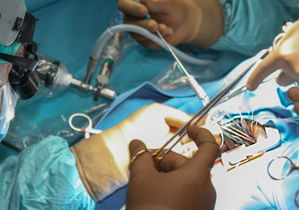
During surgical annuloplasty, the cardiac surgeon implants a specially designed support ring or partial ring, securing it to the valve tissue with sutures. The implant helps restore the normal geometry of the valve opening and improves leaflet coaptation, allowing the valve to close more effectively.
However, annuloplasty is rarely performed as a standalone procedure. In patients with degenerative mitral regurgitation, it is often combined with leaflet repair or chordal reconstruction. This combination addresses both the immediate cause of blood leakage and structural factors that may affect the long-term durability of the repair.
At Helios Heart Center Leipzig, annuloplasty using a prosthetic support ring is among the techniques used for mitral valve repair. Procedures involving isolated mitral valve disease are predominantly performed through a small incision on the right side of the chest. Cardiopulmonary bypass is used during surgery, and the effectiveness of the repair is assessed intraoperatively.
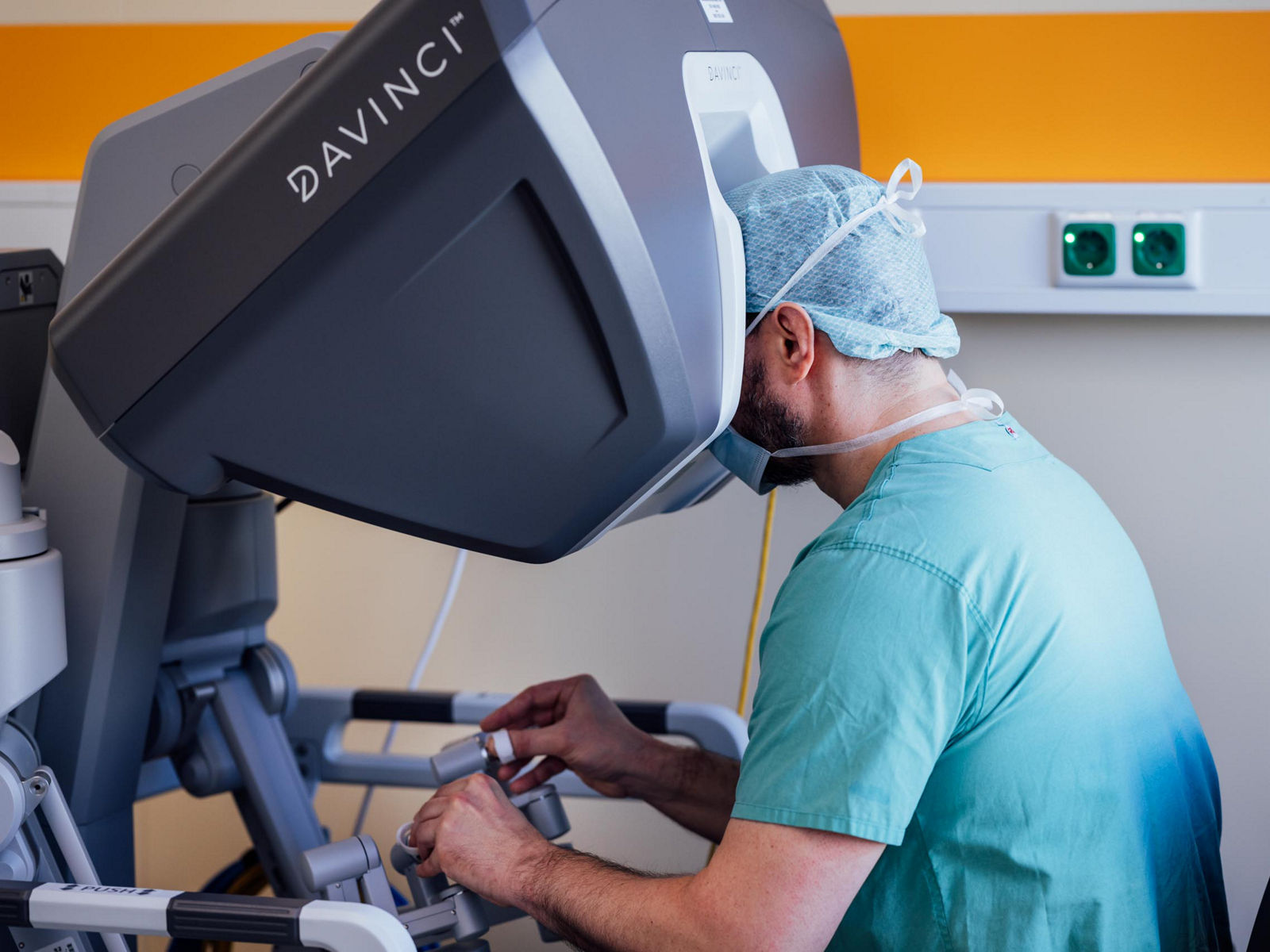
At Helios Hospital Krefeld, mitral valve repair also focuses on preserving the patient's native valve. For isolated mitral valve procedures, surgeons predominantly use a minimally invasive approach through an approximately 5–7 cm incision between the ribs on the right side of the chest, supported by three-dimensional endoscopic imaging.
Transcatheter annuloplasty: the Carillon system
In addition to surgical annuloplasty involving the implantation of a support ring, transcatheter techniques are available to reshape the mitral annulus. One such technique uses the Carillon Mitral Contour System, which can reduce mitral regurgitation without open-heart surgery.
Unlike conventional annuloplasty, in which the surgeon directly attaches a support ring to the mitral valve tissue, the Carillon system is implanted in the coronary sinus, a venous vessel running close to the mitral annulus. The catheter carrying the implant is introduced through the jugular vein in the neck. Once positioned, the device exerts tension on the surrounding structures, reshaping the mitral annulus and helping improve leaflet coaptation. Carillon is primarily used to treat functional (secondary) mitral regurgitation associated with left ventricular enlargement and changes in mitral valve geometry.
At Helios Hospital Schwerin, transcatheter annuloplasty using the Carillon system is among the available treatment options for mitral regurgitation. This technique is considered for selected patients with suitable anatomy and clinical indications.
Mitral valve leaflet repair
Mitral valve leaflet repair is performed in patients with mitral valve prolapse, degenerative changes, and other structural abnormalities that prevent the valve leaflets from closing properly. Depending on the type and extent of damage, the cardiac surgeon may remove excess tissue, reshape a leaflet, or reconstruct a damaged area.
Several established techniques are used in modern mitral valve repair:
- Triangular resection. The surgeon removes a small triangular section of damaged or excessively mobile leaflet tissue and sutures the remaining edges together. This technique is particularly suitable for localized posterior leaflet prolapse involving a relatively small area of tissue.
- Quadrangular resection. In cases of more extensive posterior leaflet prolapse, the surgeon may remove a quadrangular segment of excess tissue and reconnect the remaining sections. This is one of the classic mitral valve repair techniques developed as part of the reconstructive approach pioneered by French cardiac surgeon Alain Carpentier.
- Sliding leaflet plasty. This technique is often combined with quadrangular resection, particularly when there is significant excess tissue in the posterior leaflet. The surgeon partially detaches adjacent leaflet segments from the mitral annulus, repositions them, and reattaches them in a new position. This helps reduce posterior leaflet height, restore its shape, and lower the risk of impaired blood flow after repair.
- Folding plasty (foldoplasty). Instead of removing excess tissue, the surgeon creates a fold and secures it with sutures to reduce the height or excessive mobility of a leaflet segment. This tissue-preserving technique may be used in selected cases of mitral valve prolapse.
- Patch repair. When a leaflet has a localized defect or perforation, for example following infective endocarditis, the damaged area can sometimes be reconstructed using a patch made from biological material. The feasibility of this approach depends on the size of the defect, the condition of the surrounding tissue, and whether the infection is still active.
Modern reconstructive surgery increasingly incorporates the “respect rather than resect” principle, which emphasizes preserving native leaflet tissue rather than removing it. This approach focuses on restoring proper leaflet position and function, particularly through the use of artificial chordae tendineae. However, resectional and non-resectional techniques are not mutually exclusive. The choice depends on the valve's anatomy, and several methods may be combined in complex cases.
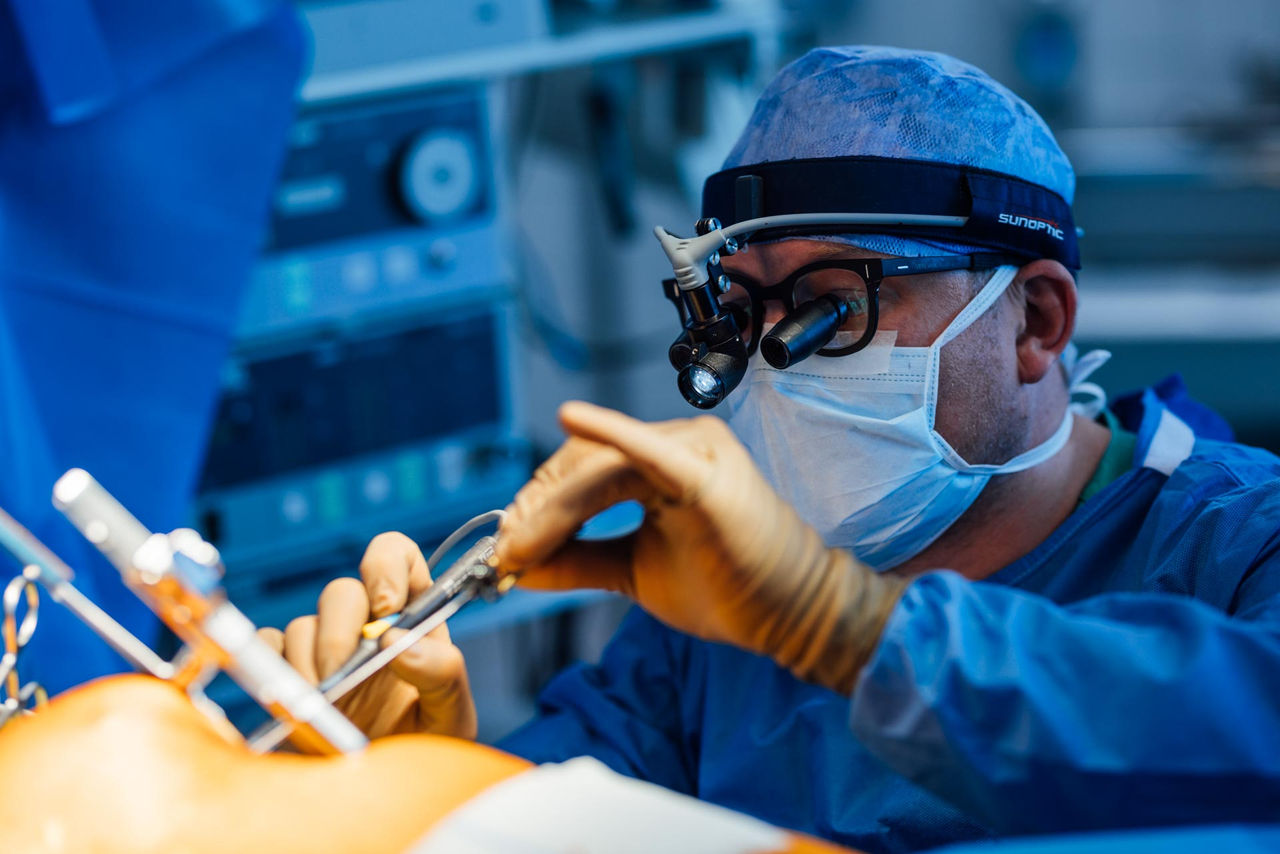
Leaflet repair is frequently combined with annuloplasty to stabilize the mitral annulus. Many reconstructive procedures can be performed using a minimally invasive approach through a small incision on the right side of the chest, supported by endoscopic imaging.
At Helios Hospital Siegburg, reconstructive mitral valve surgery is one of the cardiac surgery team's specialized areas of expertise. Whenever the valve anatomy allows for a reliable and durable result, the team aims to preserve the patient's native valve.
Helios Heart Center Leipzig has extensive experience in minimally invasive mitral valve repair, including complex combined procedures. Depending on the characteristics of the valve disease, surgeons may combine leaflet reconstruction with chordal repair and annuloplasty.
Chordal repair and reconstruction
The chordae tendineae are thin, strong fibrous cords that connect the mitral valve leaflets to the papillary muscles of the left ventricle. They keep the leaflets in the correct position during heart contraction and prevent them from moving excessively into the left atrium.
Degenerative changes can cause the chordae to become elongated or rupture. This may lead to mitral valve leaflet prolapse and, in some cases, the sudden onset of severe mitral regurgitation.
One modern reconstructive technique is artificial chordae implantation (neochordae implantation). The surgeon uses thin sutures made from a biocompatible material, usually polytetrafluoroethylene (PTFE), to replace damaged chordae. These artificial chordae are attached to the appropriate structures of the mitral valve apparatus, and their length is adjusted to ensure proper leaflet coaptation.
Unlike valve replacement, this procedure preserves the natural anatomy of the mitral valve and its connection to the left ventricle. When necessary, neochordae implantation is combined with annuloplasty.
Helios Heart Center Leipzig uses reconstructive techniques involving the implantation of artificial chordae tendineae and an annuloplasty ring. In patients with suitable anatomy, the procedure is performed using a minimally invasive approach through the right side of the chest. This technique restores valve function without the need to implant a mechanical or biological valve prosthesis.
Transcatheter mitral valve repair — TEER
TEER (transcatheter edge-to-edge repair) is a minimally invasive treatment for mitral regurgitation that improves valve leaflet coaptation without open-heart surgery. Unlike surgical repair, in which a cardiac surgeon directly reconstructs damaged valve structures, TEER involves implanting a small device that connects portions of the anterior and posterior mitral valve leaflets.
The procedure is performed using a catheter inserted through the femoral vein in the groin. The physician advances the catheter toward the heart and reaches the mitral valve by crossing the interatrial septum. Under three-dimensional transesophageal echocardiographic and fluoroscopic guidance, the device grasps the leaflets at the site of incomplete closure and brings them closer together. This reduces the opening through which blood flows backward into the left atrium. The patient's native valve is preserved, and cardiopulmonary bypass is not required.
Two main systems are used for TEER: MitraClip and PASCAL. Both serve the same purpose but differ in implant design and the way they interact with the valve leaflets.
MitraClip
MitraClip is a technique that connects the valve leaflets using a specialized clip. The device consists of a small metallic clip with two movable grasping arms. Once delivered to the mitral valve, the clip is positioned between the anterior and posterior leaflets. The physician grasps portions of both leaflets and closes the device, securing them together. This improves leaflet coaptation during heart contraction and reduces the amount of blood flowing backward.
Modern generations of MitraClip are available in several sizes, with different widths and lengths of the grasping arms. This allows physicians to select a device based on leaflet thickness, length, and mobility, as well as the location of the regurgitant jet. The latest generations allow the anterior and posterior leaflets to be grasped either simultaneously or independently, helping achieve precise positioning and fixation.
If a single clip does not sufficiently reduce regurgitation, additional devices may be implanted when the valve anatomy is suitable. Throughout the procedure, specialists assess not only the reduction in backward blood flow but also the remaining valve opening area to avoid significant mitral valve narrowing.
PASCAL
PASCAL uses the same TEER principle of bringing the anterior and posterior mitral valve leaflets closer together. However, its design differs from MitraClip.
The main distinguishing feature of PASCAL is an additional central spacer, positioned between two broad, flexible grasping elements known as paddles. When the device is secured to the leaflets, the central spacer occupies part of the gap between them at the site of incomplete closure. As a result, regurgitation is reduced not only by bringing the leaflets together but also by filling part of the space through which blood leaks backward.
The broad, curved paddles of the PASCAL system are designed to distribute force across the leaflet tissue. Additional clasps secure each leaflet and allow independent leaflet grasping. The device can also be temporarily elongated during delivery and repositioning, making it easier to navigate around the chordae tendineae.
The system is available in different implant configurations, including narrower options designed for specific anatomical characteristics of the mitral valve.
At Helios Hospital Erfurt, Helios Hospital Schwerin, Helios Hospital Krefeld, and Helios Heart Center Leipzig, TEER is among the treatment options for mitral regurgitation. These include MitraClip and PASCAL technologies, with the fifth-generation MitraClip system also used at selected Helios centers.
Mitral valve replacement techniques
Mitral valve replacement (MVR) is a procedure in which a damaged valve is replaced with an artificial prosthesis. Unlike mitral valve repair, which aims to restore the patient's own valve leaflets and chordae tendineae, replacement makes it possible to replace a valve that can no longer be reliably repaired.
During surgical replacement, the affected valve leaflets are removed completely or partially, while the chordae tendineae and other components of the subvalvular apparatus are preserved whenever possible. These structures connect the valve to the papillary muscles and help maintain the normal geometry of the left ventricle.
Mitral valve replacement may be necessary in cases of severe leaflet calcification and deformation, severe stenosis, extensive tissue destruction caused by infective endocarditis, and complex forms of mitral regurgitation when repair would not provide a durable result. The replacement technique and type of prosthesis are selected based on the anatomy of the heart, the patient's age, coexisting conditions, and the feasibility of long-term anticoagulant therapy.
Types of mitral valve prostheses
Two main types of prostheses are used for mitral valve replacement: mechanical and biological valves. Both are designed to restore one-way blood flow, but they differ in construction, durability, and the medication required after implantation.
Mechanical mitral valves are made from highly durable biocompatible materials, primarily pyrolytic carbon. The most common modern design is a bileaflet prosthesis: two movable leaflets open as blood enters the left ventricle and close when it contracts.
Mechanical valves are highly resistant to wear and are designed to function for a long time without structural deterioration of their leaflets. They are therefore often considered for younger patients for whom reducing the likelihood of repeat surgery due to prosthetic valve deterioration is particularly important.
The main characteristic of a mechanical valve is the need for lifelong anticoagulant therapy with vitamin K antagonists, such as phenprocoumon or warfarin. These medications prevent blood clots from forming on the artificial surfaces of the valve. Treatment is monitored through regular measurement of the international normalized ratio (INR). For most mechanical prostheses in the mitral position, the target INR is 3.0, with a therapeutic range of 2.5–3.5; a physician may prescribe a higher target value if additional risk factors are present. Direct oral anticoagulants, including apixaban and rivaroxaban, are not used for mechanical valves.
Biological mitral valves are made from specially treated animal tissues, most commonly porcine heart valves or bovine pericardium. The leaflets are attached to a supporting frame that helps maintain the shape of the prosthesis and ensures its fixation within the heart.
“The lifespan of a biological heart valve cannot be predicted with certainty,” — Prof. Dr. med. Michael A. Borger, Director of the University Department of Cardiac Surgery, Helios Heart Center Leipzig.
Bioprosthetic valves have lower thrombogenicity and therefore generally do not require lifelong anticoagulation solely because of the implanted valve. However, after surgical implantation of a biological mitral prosthesis, anticoagulant therapy is prescribed for at least the first three months. In patients with atrial fibrillation or other independent indications, long-term treatment may be necessary.
The main limitation of biological valves is the gradual deterioration of their tissues. Over time, the leaflets may become calcified, thickened, or damaged, leading to recurrent stenosis or mitral regurgitation. The durability of a bioprosthesis depends on the model, the patient's age, and individual factors: structural degeneration generally develops more rapidly in younger patients.
The choice between a mechanical and a biological valve is not determined by age alone. The cardiac surgeon considers life expectancy, bleeding risk, the ability to undergo regular INR monitoring, coexisting conditions, and patient preferences. Biological valves are more often selected for patients at increased risk of anticoagulation-related complications and for women planning pregnancy. Mechanical prostheses may be preferable when implant durability is particularly important.
Surgical mitral valve replacement — SMVR
Surgical mitral valve replacement (SMVR) is a procedure in which a cardiac surgeon directly removes damaged valve tissue and replaces it with a mechanical or biological prosthesis. The operation is performed under general anesthesia using a heart-lung machine, which temporarily maintains blood circulation while the surgeon operates on the heart.
After accessing the mitral valve, the surgeon assesses the condition of the leaflets, chordae tendineae, papillary muscles, and mitral annulus. The affected tissues are removed to the extent necessary for prosthesis implantation. The surgeon then selects the appropriate prosthesis size and secures it to the mitral annulus using special sutures. It is important not only to ensure secure fixation but also to avoid narrowing the valve opening or obstructing blood flow within the left ventricle.
One of the key technical approaches in modern surgical replacement is mitral valve replacement with preservation of the subvalvular apparatus (chordal-sparing MVR). In the past, the leaflets and chordae were often removed completely during valve replacement. Today, whenever tissue conditions permit, surgeons aim to preserve their connection to the papillary muscles, as this contributes to maintaining the shape and contractile function of the left ventricle.
Several variations of this technique are used depending on the anatomy. With posterior leaflet preservation, the posterior leaflet and its attached chordae are retained. With bileaflet preservation, surgeons aim to preserve the subvalvular structures of both leaflets. In some cases, the remaining tissues are repositioned or secured so that they do not interfere with the movement of the new prosthesis or obstruct blood flow out of the left ventricle.
Preservation of the chordae is not always possible. For example, in infective endocarditis, the surgeon must completely remove infected and destroyed tissues. In cases of severe mitral annular calcification, additional techniques may be required to prepare the implantation site, including limited removal of calcium deposits, tissue reinforcement, or reconstruction of individual sections of the annulus using a pericardial patch. These measures form part of surgical valve replacement and are selected according to the specific valve pathology.
SMVR can be performed through either a traditional sternotomy or a minimally invasive approach. In conventional surgery, the surgeon divides the sternum to obtain a wide view of the heart. This approach may be necessary in cases of complex anatomical changes, repeat operations, or when other cardiac surgical procedures must be performed simultaneously.
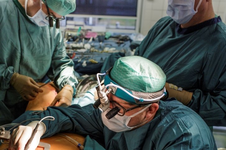
Minimally invasive valve replacement uses a small incision on the right side of the chest, known as a mini-thoracotomy. Surgical instruments are introduced through this incision, and an endoscopic camera providing three-dimensional images may be used for visualization. The heart-lung machine is often connected through blood vessels in the groin. Despite the smaller incision, this remains a full surgical valve replacement procedure rather than a catheter-based implantation.
A minimally invasive approach avoids complete division of the sternum and, in suitable patients, may reduce chest trauma, postoperative pain, and recovery time. However, its use depends on the condition of the blood vessels, cardiac anatomy, the extent of calcification, and the scope of the planned operation.
At Helios Hospital Siegburg, mitral valve procedures are performed through a right-sided mini-thoracotomy with endoscopic visualization when appropriate. At Helios Hospital Krefeld, minimally invasive approaches also play an important role in heart valve surgery. The choice of surgical access does not limit the possibility of implanting a mechanical or biological prosthesis; it is determined by the characteristics of the disease and the safety of the procedure.
After securing the prosthesis, the surgeon checks its function using transesophageal echocardiography. This examination makes it possible to confirm proper leaflet movement, assess blood flow through the valve, and rule out significant leakage between the prosthesis and the surrounding tissues.
Transcatheter mitral valve replacement — TMVR
Transcatheter mitral valve replacement (TMVR), also known as transcatheter mitral valve implantation (TMVI), allows a new valve to be implanted without conventional surgery involving direct removal of the damaged mitral valve.
The prosthesis is delivered to the heart in a compressed state using a catheter system, positioned correctly, and expanded. Unlike TEER repair using MitraClip or PASCAL, in which the patient's own leaflets are brought closer together to reduce regurgitation, TMVR involves implanting a new valve that takes over the function of the damaged valve.
Different access routes may be used to deliver the prosthesis. With the transseptal approach, a catheter is introduced through the femoral vein and advanced into the left atrium through the interatrial septum. This method does not require opening the chest. With the transapical approach, the catheter is introduced directly into the region of the left ventricular apex through a small chest incision. This approach is more invasive, although it avoids conventional surgery involving complete division of the sternum.
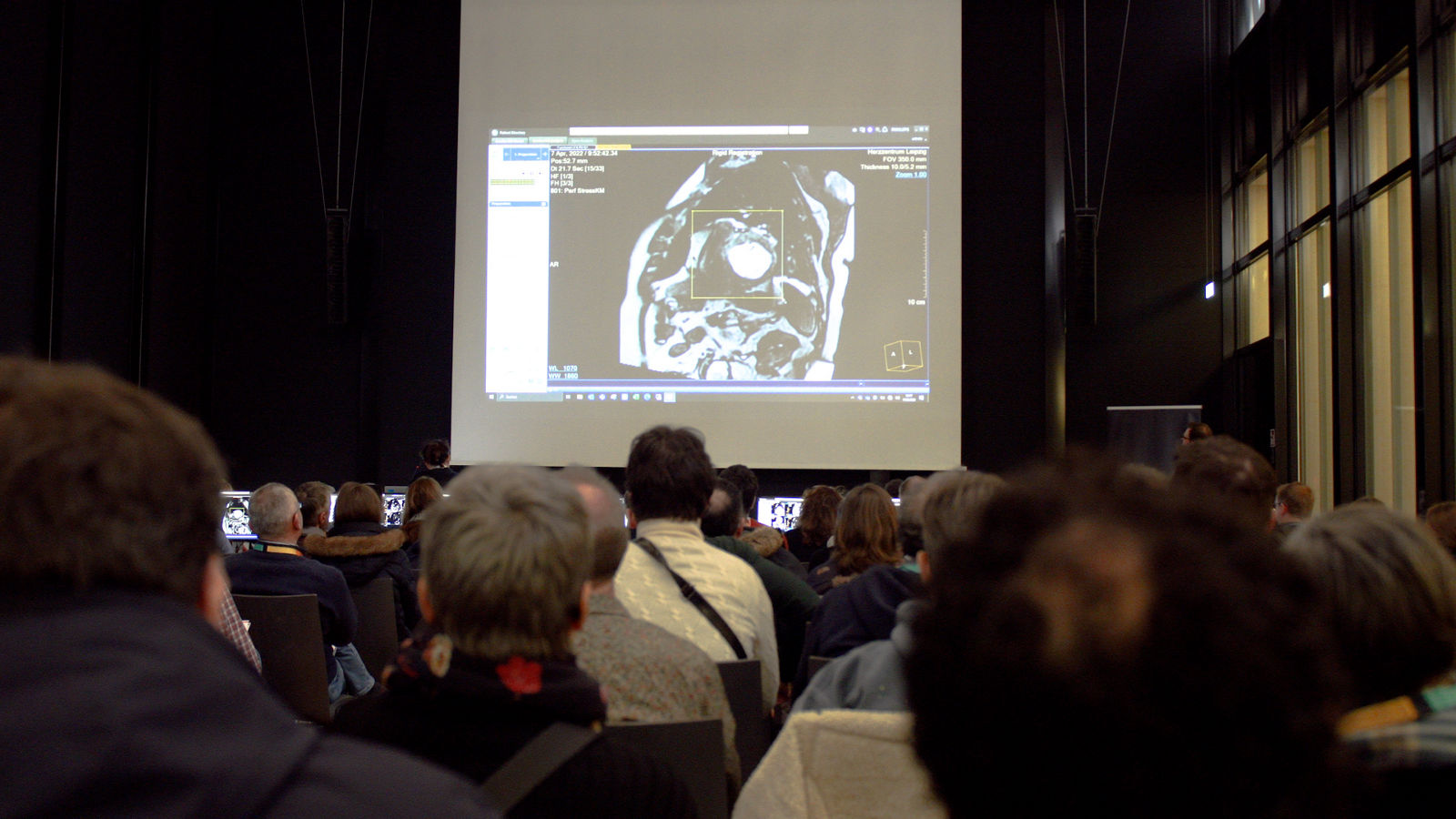
TMVR requires particularly precise planning. The mitral annulus has a complex shape and may be significantly dilated or calcified. In addition, an incorrectly positioned prosthesis can obstruct the left ventricular outflow tract — the pathway through which blood enters the aorta. Therefore, echocardiography and cardiac computed tomography with three-dimensional reconstruction are performed before the procedure to assess the dimensions of the valve apparatus, the anticipated implant position, and the risk of complications.
Today, transcatheter valve replacement is used in several clinical situations.
Valve-in-valve: replacement of a deteriorated bioprosthesis
Mitral valve-in-valve (ViV) is used in patients who previously underwent surgical implantation of a biological mitral valve that has subsequently lost its normal function. This may result from calcification and restricted leaflet mobility, leaflet damage, or the development of recurrent mitral regurgitation.
During a valve-in-valve procedure, the old bioprosthesis is generally not removed. A new valve is delivered through a catheter and expanded inside the previously implanted prosthesis. The frame of the existing valve serves as a support for the new implant, ensuring its fixation.
The procedure is most commonly performed transseptally through the femoral vein, without opening the chest or using a heart-lung machine. The position of the prosthesis is monitored using fluoroscopy and transesophageal echocardiography.
One of the main advantages of valve-in-valve is the possibility of avoiding repeat open-heart surgery, particularly in patients at increased surgical risk. However, the technique is not suitable for all bioprostheses. The physician must know the exact model and internal diameter of the previously implanted valve: an excessively small diameter may result in increased resistance to blood flow after implantation of the new prosthesis.
Valve-in-ring: valve implantation after previous repair
Mitral valve-in-ring (ViR) is used in a different situation: the patient's own mitral valve was previously repaired with the implantation of an annuloplasty ring, but severe recurrent regurgitation or stenosis subsequently developed.
Instead of repeat surgery, a biological valve prosthesis can be implanted inside the previously placed ring in selected cases. The new valve expands within the mitral orifice and takes over the function of the damaged valve.
In terms of delivery, valve-in-ring is similar to valve-in-valve and can also be performed transseptally through the femoral vein. However, prosthesis fixation is technically more challenging: annuloplasty rings vary in shape, size, and rigidity, and some models do not form a completely closed frame.
Before the procedure, specialists review the records from the previous operation, identify the type of ring, and perform computed tomography. This is necessary to assess whether the new valve can be securely fixed and to evaluate the risks of valve displacement, leakage around the implant, and obstruction of the left ventricular outflow tract.
Transcatheter replacement of the native mitral valve — native-valve TMVR
A separate approach is native-valve TMVR, in which a new prosthesis is implanted directly in the region of the patient's own mitral valve rather than inside a previously implanted bioprosthesis or annuloplasty ring.
This is a technically more complex procedure. The native mitral annulus, particularly in patients with mitral regurgitation, is often dilated and lacks the rigid support needed to secure a conventional transcatheter prosthesis. For this reason, specialized systems with dedicated fixation mechanisms are being developed for these procedures.
One example is Tendyne, a self-expanding biological prosthesis with a dual-contour design that adapts to the shape of the mitral orifice. The system is implanted through a transapical approach and held in place by a special anchoring mechanism in the region of the left ventricular apex. This technique allows valve implantation without a conventional sternotomy, although it requires a small surgical incision in the chest.
Another example is SAPIEN M3, a system designed for transseptal implantation. First, a special supporting structure that interacts with the native valve apparatus is implanted, and then a biological prosthesis is expanded inside it. This approach allows catheter-based access through the femoral vein.
Direct transcatheter replacement of the native mitral valve is currently used much more selectively than surgical valve replacement. Its use depends on cardiac anatomy, clinical indications, the availability of the specific system, and the experience of the specialized center. These technologies cannot be considered a universal alternative to surgery.
In selected patients with severe mitral annular calcification, valve-in-MAC may also be considered. This involves transcatheter implantation of a prosthesis supported by calcified tissue. However, the procedure carries an increased risk of valve displacement, leakage around the prosthesis, and left ventricular outflow tract obstruction. It therefore requires particularly careful patient selection and is not a standard replacement option for most patients.
Percutaneous balloon mitral commissurotomy
Percutaneous balloon mitral commissurotomy (PMC) is neither a mitral valve repair nor a replacement procedure. However, it is an important standalone treatment for mitral stenosis that allows the narrowed valve opening to be widened and blood flow to be restored without implanting an artificial prosthesis.
The technique is primarily used for rheumatic mitral stenosis. In this condition, the commissures — the areas where the anterior and posterior valve leaflets meet — become fused. As a result, the leaflets lose mobility, the valve opening narrows, and blood flow from the left atrium to the left ventricle becomes restricted.
The procedure is performed through the femoral vein. The physician introduces a catheter, advances it through the interatrial septum into the left atrium, and then passes it through the narrowed mitral valve. A specialized Inoue balloon is often used to widen the opening. It is gradually inflated under fluoroscopic and echocardiographic guidance. The pressure of the balloon separates the fused commissures, increasing the valve opening area and reducing resistance to blood flow. The procedure does not require opening the chest or using a heart-lung machine.
Suitable valve anatomy is the key requirement for successful commissurotomy. The best results are achieved when there is significant commissural fusion, the leaflets remain sufficiently mobile, and substantial calcification is absent. Before the procedure, transthoracic and, when necessary, transesophageal echocardiography are performed to assess valve anatomy, the severity of stenosis, and to rule out a blood clot in the left atrium.
Balloon commissurotomy is not performed in patients with a thrombus in the left atrium, more than mild mitral regurgitation, severe calcification, or no commissural fusion. In such cases, surgical treatment is considered. The main procedural risk is damage to the valve leaflets, which may cause or worsen mitral regurgitation.
When the procedure is successful, pressure in the left atrium and pulmonary vessels decreases, which can relieve shortness of breath and improve exercise tolerance. However, the valve may narrow again over time, a condition known as restenosis. Therefore, regular cardiology follow-up with echocardiographic monitoring is necessary after commissurotomy. When the result is stable and the patient has no symptoms, examinations are generally performed annually, or more frequently if restenosis develops or symptoms return.
Life after mitral valve repair or replacement
After successful mitral valve repair or replacement, patients usually experience less shortness of breath, fatigue, and other symptoms associated with impaired blood flow. As heart function improves, physical activity becomes easier, allowing patients to return to work and their usual activities. However, long-term well-being depends not only on the outcome of the procedure but also on the condition of the heart muscle, the presence of arrhythmias, and other underlying diseases.
Life after treatment also depends largely on whether the patient's own valve was repaired or a prosthesis was implanted. This determines the medication required and the long-term follow-up schedule.
Recovery and rehabilitation
After surgical mitral valve repair or replacement, rehabilitation begins in the hospital. Patients gradually resume walking and everyday activities, perform breathing exercises, and then progress to supervised physical training. Cardiac rehabilitation helps restore endurance, safely increase physical activity, and reduce the risk of postoperative complications.
The main recovery period after surgery usually takes 4–8 weeks, although complex operations and coexisting conditions may require more time. After a sternotomy, additional time is needed for the breastbone to heal. Recovery is often faster after minimally invasive surgery because there is less damage to the tissues of the chest.
After transcatheter procedures, such as TEER or transseptal TMVR, patients generally need less time to return to everyday activities. However, even after a technically successful procedure, patients with heart failure or reduced left ventricular function may still require comprehensive cardiac rehabilitation.
Living with a repaired valve or valve prosthesis
After mitral valve repair, patients generally do not require permanent anticoagulant therapy unless there are other indications. Antithrombotic medications may be prescribed during the first few months after the procedure, while patients with atrial fibrillation or an increased risk of thromboembolism may need long-term treatment. Ongoing monitoring of the repaired valve is important because mitral regurgitation may recur over time.
A mechanical mitral valve prosthesis is designed for long-term function and is highly resistant to wear. However, blood clots can form on its artificial surfaces, so patients must take vitamin K antagonists, such as phenprocoumon or warfarin, for life. The effectiveness of treatment is monitored using the international normalized ratio (INR). For most mechanical mitral valves, the target INR is 3.0, with a therapeutic range of 2.5–3.5. Testing is more frequent at the beginning of treatment, and once the values stabilize, the intervals between measurements can be extended according to the physician's recommendations.
A biological mitral valve prosthesis generally does not require lifelong anticoagulation unless there are other indications. After surgical implantation of a bioprosthesis, anticoagulant therapy is prescribed for at least three months, while after transcatheter implantation, it is usually continued for at least six months. However, biological tissues gradually age: over time, the leaflets may become calcified or lose mobility, leading to recurrent stenosis or mitral regurgitation. Therefore, patients need regular assessments of prosthetic valve function even when they feel well.
What examinations are needed and how often?
The main method of monitoring after mitral valve treatment is echocardiography (cardiac ultrasound). It allows physicians to assess the function of the repaired valve or prosthesis, detect recurrent regurgitation, narrowing of the valve opening, and changes in left ventricular function. The first postoperative examination also provides a baseline for comparison with subsequent results.
Patients with heart failure, arrhythmias, residual mitral regurgitation, or suspected prosthetic valve dysfunction require more frequent examinations. Additional tests may include an electrocardiogram (ECG) or Holter monitoring to assess heart rhythm, blood tests to monitor treatment, and, when necessary, transesophageal echocardiography or cardiac computed tomography.
Daily life and prevention of complications
After completing rehabilitation, most patients can lead active lives, gradually increasing physical activity according to their cardiologist's recommendations. It is important to continue prescribed treatment and attend regular follow-up examinations, even if symptoms have completely disappeared.
Particular attention is given to preventing infective endocarditis, an infection that can affect both the patient's own valve and an implanted prosthesis. Patients should maintain good oral hygiene, visit their dentist regularly, and inform healthcare professionals about their previous valve procedure. Patients with a prosthetic valve or implanted material used for valve repair require antibiotic prophylaxis before certain dental procedures.
If shortness of breath, swelling, palpitations, or severe fatigue return after treatment, patients should contact their cardiologist rather than wait for their next scheduled appointment. Timely diagnosis helps identify possible changes in valve function and preserve the benefits achieved through treatment.
Mitral valve treatment at Helios hospitals
Helios hospitals in Germany offer advanced diagnostic and treatment methods for mitral valve diseases, ranging from surgical repair to valve replacement and transcatheter interventions. Patients receive medical care in specialized cardiology and cardiac surgery departments equipped with advanced technology for precise diagnostics, complex surgical procedures, and postoperative monitoring.
Treatment involves a multidisciplinary Heart Team consisting of experienced cardiologists, cardiac surgeons, cardiac imaging specialists, and anesthesiologists. Joint assessment of echocardiography, computed tomography, and other examination results allows the team to select the most appropriate treatment strategy based on valve anatomy, the condition of the heart muscle, and individual risks. Priority is given to preserving the patient's native valve whenever repair can provide a reliable long-term result.
“In complex heart valve diseases, all relevant factors are assessed by a specialized Heart Team,” — Prof. Dr. med. Michael A. Borger, Director of the University Department of Cardiac Surgery, Helios Heart Center Leipzig.
Particular attention is paid to patient safety and comfort at every stage of treatment. Before the procedure, patients undergo a comprehensive examination and specialist consultations, while modern anesthesia techniques and vital function monitoring are used during surgery. After surgical treatment, patients initially receive intensive medical monitoring and are then transferred to a regular hospital room once their condition stabilizes. The medical team monitors heart function, tissue healing, the effectiveness of prescribed medications, and the recovery of physical activity.
Hospital stays at Helios are organized with the needs of international patients in mind. Comfortable rooms, attentive medical staff, and well-coordinated examinations and procedures help patients focus on their treatment and recovery. When necessary, an individualized cardiac rehabilitation and follow-up plan is developed after surgery.
The Helios International team supports international patients throughout the organizational stages of treatment in Germany: helping them select a suitable hospital, arrange a review of their medical records, obtain a preliminary treatment plan, and coordinate hospital admission. The team also provides assistance with communication with the hospital and travel preparations.
The combination of experienced cardiology and cardiac surgery teams, modern technologies, and comprehensive postoperative care enables Helios to provide an individualized approach to mitral valve disease treatment — from initial diagnosis to recovery and long-term follow-up.
If you are interested in arranging diagnosis or treatment at one of the Helios hospitals, you can submit a request using the contact form on our website or contact us via the email address provided at the top of the website.
Learn more about arranging treatment at Helios hospitals ⟶
Do you need more information about Helios Hospitals or want to schedule your treatment?