Aortic valve replacement and repair
- Causes of aortic valve replacement
- Aortic valve repair and reconstruction
- Aortic valve replacement methods
- Choosing the treatment method and preparing for surgery
- Rehabilitation and life after surgery
- Aortic valve replacement and repair at Helios hospitals
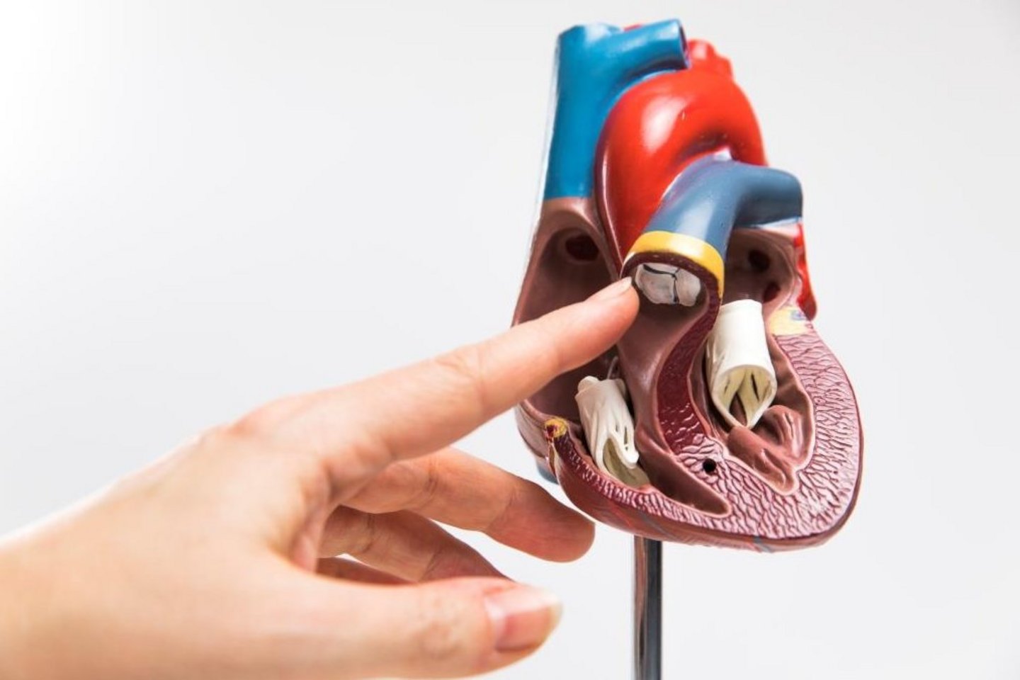
The aortic valve is one of the heart’s four valves, located between the left ventricle and the aorta, the largest artery in the body. It allows oxygen-rich blood to flow from the heart to the organs and tissues while preventing it from flowing backward. When the valve does not function properly, the workload on the heart increases, which can eventually lead to heart failure. Depending on the type and severity of the condition, treatment may include medication to relieve symptoms and manage associated conditions. However, severe valve disease may require surgical replacement or repair.
Aortic valve diseases pose a significant global health challenge, particularly among older adults. According to estimates from the international JACC study (Global Burden of Cardiovascular Diseases), non-rheumatic heart valve diseases caused approximately 191,000 deaths worldwide in 2023. Of these, 149,400 deaths (around 78%) were attributed to calcific aortic valve disease.
Let's take a closer look at the conditions that may require aortic valve replacement.
Causes of aortic valve replacement
Aortic valve dysfunction can result from age-related changes, congenital abnormalities, infections, and diseases of the aorta. Depending on the type of damage, the valve may become narrowed, restricting normal blood flow (aortic stenosis), or fail to close completely, allowing blood to flow backward into the left ventricle (aortic regurgitation).
The main causes of aortic valve dysfunction include:
- Degenerative changes and calcification. With age, calcium deposits may accumulate on the valve leaflets, causing them to thicken and lose their elasticity and mobility. This is the most common cause of aortic stenosis in older adults.
- Congenital aortic valve defects. The most common is a bicuspid aortic valve, which has two leaflets instead of the usual three. This condition increases the risk of developing aortic stenosis or regurgitation at an earlier age.
- Infective endocarditis. A bacterial or, less commonly, fungal infection of the heart's inner lining can affect the valve, damage its leaflets, and cause acute aortic regurgitation.
- Rheumatic valve disease. This develops as a result of rheumatic fever, which is associated with an immune response to a streptococcal infection. It can cause thickening, deformation, and fusion of the valve leaflets, leading to stenosis, regurgitation, or a combination of both.
- Aortic root dilation and ascending aortic aneurysm. When the aortic root becomes enlarged, the valve leaflets may no longer close properly, even if their structure remains intact. This can lead to aortic regurgitation.
- Leaflet degeneration and prolapse. Changes in connective tissue, stretching, or abnormal positioning of the valve leaflets may prevent them from closing completely, allowing blood to flow backward.
When is aortic valve replacement or repair necessary?
Not all of these conditions require surgery. In cases of mild or moderate valve dysfunction, regular cardiological monitoring and medication to manage associated conditions are often sufficient. The decision to perform surgery depends primarily on the severity of valve dysfunction, the presence of symptoms, and the condition of the heart muscle.
The most common indication for aortic valve replacement is severe aortic stenosis, primarily caused by age-related calcification or a congenital bicuspid aortic valve. Intervention is usually necessary when symptoms such as shortness of breath, chest pain, or fainting occur. In certain cases, intervention may also be recommended for asymptomatic patients, for example, when the pumping function of the left ventricle is reduced.
Severe aortic regurgitation may also require surgical treatment, particularly when symptoms develop, the left ventricle becomes enlarged, or its function deteriorates. Depending on the condition of the valve leaflets, either valve replacement or repair may be possible.
Special attention is required in situations where urgent or emergency surgery may be necessary:
- Infective endocarditis — in cases of severe valve regurgitation accompanied by heart failure, uncontrolled infection, or other serious complications.
- Acute ascending aortic dissection — when the aortic root is affected and valve function is impaired, emergency surgery may be required to preserve or replace the valve.
- Acute severe aortic regurgitation — for example, due to leaflet destruction caused by endocarditis or aortic dissection.
In cases of aortic root dilation without significant damage to the valve leaflets, valve-sparing surgery may be preferred in suitable patients. However, if the leaflets are irreversibly damaged or severely calcified, valve replacement is more often required.
Aortic valve repair and reconstruction
Aortic valve repair and reconstruction aim to restore normal valve function without implanting an artificial valve prosthesis. Depending on the type of damage, the surgeon may repair the patient's existing valve leaflets, create new ones from the patient's own tissue, or reconstruct the aortic root while preserving the valve. The main advantage of these procedures is the ability to preserve natural valve function and avoid the lifelong anticoagulant therapy required with mechanical valve replacement.
Several techniques are available for aortic valve repair and reconstruction. The choice depends on the condition of the valve leaflets, the anatomy of the aortic root, and the underlying cause of the disease. Let's take a closer look at the main surgical techniques.
Aortic valve leaflet repair
Aortic valve leaflet repair is a surgical technique used to restore the patient's own valve leaflets without completely replacing them with a prosthesis. The aim of the procedure is to correct leaflet deformities and damage, ensuring proper closure and preventing blood from flowing backward into the left ventricle.
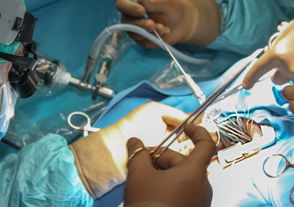
The operation is performed under general anesthesia using a heart-lung machine (cardiopulmonary bypass). Depending on the type of damage, the surgeon may use different techniques:
- Leaflet plication — excess or stretched leaflet tissue is folded and secured with surgical sutures to correct its shape and the height of its free edge.
- Free-edge reinforcement — a thin surgical suture is placed along the edge of the leaflet to restore proper tension and correct sagging (prolapse).
- Closure of small defects — small holes or tears in the valve leaflets may be closed with surgical sutures in suitable cases.
- Pericardial patch repair — larger localized defects may be repaired using a patch made from the patient's own pericardium (the membrane surrounding the heart) or another suitable biological material.
Leaflet repair is primarily performed in patients with aortic regurgitation caused by leaflet prolapse, stretching, or localized damage. It may also be used for certain congenital abnormalities, including bicuspid aortic valves.
The main advantage of this technique is the preservation of the patient's own valve, eliminating the need for lifelong anticoagulant therapy solely because of the procedure. However, repair may not be possible in cases of severe calcification, extensive leaflet destruction, or significant deformity. There is also a risk of recurrent aortic regurgitation over time, which may require further intervention.
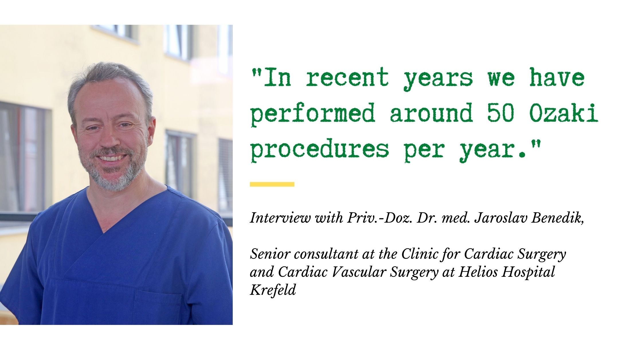
Ozaki procedure
The Ozaki procedure is a method of aortic valve reconstruction in which damaged valve leaflets are replaced with new ones made from the patient's own tissue. Unlike conventional leaflet repair, the surgeon does not restore the existing leaflets but creates new ones while preserving the natural structure of the valve annulus, without implanting a mechanical or biological valve prosthesis.
The procedure is performed under general anesthesia using a heart-lung machine. The surgeon takes a small section of the pericardium — the membrane surrounding the heart — and treats it with a special solution to improve the tissue's strength and stability. Using templates, new leaflets are then cut from the prepared tissue to match the dimensions of the patient's valve. The damaged leaflets are removed, and the newly created pericardial leaflets are sutured to the valve annulus, restoring normal valve opening and closure.
The main advantage of the procedure is the use of the patient's own tissue. However, the technique is complex, and the long-term durability of the reconstructed leaflets is less well established than that of conventional valve prostheses. Over time, aortic stenosis or regurgitation may recur, potentially requiring another operation.
Valve-sparing aortic root surgery
Valve-sparing aortic root surgery refers to a group of surgical procedures designed to repair an enlarged or damaged aortic root while preserving the patient's own aortic valve. These procedures are primarily performed in patients with aortic root aneurysms and associated aortic regurgitation, provided that the valve leaflets are sufficiently healthy to be preserved.
Unlike conventional aortic valve replacement, the surgeon replaces the affected section of the aorta with a synthetic vascular graft while preserving the patient's native valve leaflets. The two main techniques are the David and Yacoub procedures.
David procedure
The David procedure, also known as aortic valve reimplantation, involves replacing the enlarged aortic root and reimplanting the patient's own aortic valve inside a vascular graft. The operation is performed under general anesthesia using a heart-lung machine.
The surgeon removes the enlarged sections of the aortic root while preserving the valve leaflets. The valve is then positioned inside a tubular synthetic graft and secured with sutures. The graft supports the valve annulus and restores the normal geometry of the aortic root. The coronary arteries, which originate from the aortic root, are reattached to the graft.
The main advantage of this technique is the preservation of the patient's own valve while reinforcing the valve annulus, reducing the risk of further dilation. The procedure is particularly suitable for patients with aortic root aneurysms, including some individuals with inherited connective tissue disorders such as Marfan syndrome. However, valve preservation may not be possible if the leaflets are severely damaged or calcified.
Yacoub procedure
The Yacoub procedure, also known as aortic root remodeling, also involves replacing an enlarged aortic root while preserving the patient's own valve, but uses a different reconstruction technique.
During the operation, the surgeon removes the enlarged sections of the aortic root and shapes the vascular graft to recreate the natural sinuses of Valsalva — the dilated portions of the aorta located just above the valve leaflets. The prepared graft is sutured to the remaining aortic root tissue, preserving the native valve and its natural mobility. The coronary arteries are then reattached to the graft.
The main advantage of this technique is the restoration of the aortic root's anatomy while preserving natural valve function. However, unlike the David procedure, the classic Yacoub technique does not provide the same degree of valve annulus stabilization. Therefore, patients with annular dilation may require additional annuloplasty — a surgical procedure to reinforce the valve annulus.
Aortic valve replacement methods
Aortic valve replacement involves implanting a new valve to replace the damaged one, restoring normal blood flow and reducing the workload on the heart. Modern cardiac surgery offers several replacement techniques that differ in how the procedure is performed and the type of valve used. Before exploring the main replacement methods, let's take a closer look at the types of prosthetic valves available and how they differ.
Biological and mechanical aortic valves
Two main types of prosthetic valves can be used for aortic valve replacement: biological and mechanical. They differ in their materials, durability, and the need for medication after surgery.
-
Biological valves are primarily made from animal tissue (porcine or bovine). Their main advantage is that they do not require lifelong anticoagulant therapy solely because of the implanted valve. However, biological prostheses gradually wear out and may eventually need to be replaced. They are generally preferred for older patients and people for whom long-term anticoagulant therapy is contraindicated.
-
Mechanical valves are made from highly durable synthetic materials, particularly pyrolytic carbon. They are designed to last for decades. Their main disadvantage is the need for lifelong anticoagulant therapy (usually warfarin or another vitamin K antagonist) to prevent blood clots, which increases the risk of bleeding. These valves are more commonly considered for younger patients with a longer life expectancy.
“The durability of a biological heart valve cannot be predicted with certainty for every patient. Factors such as the patient's age, the valve's position, and the type of prosthesis used all play a role.” — Prof. Michael A. Borger, MD, PhD, Medical Director and Director of the University Department of Cardiac Surgery at the Leipzig Heart Center.
The choice between a biological and mechanical valve depends on the patient's age, underlying medical conditions, bleeding risk, and individual preferences.
TAVI/TAVR — transcatheter aortic valve replacement
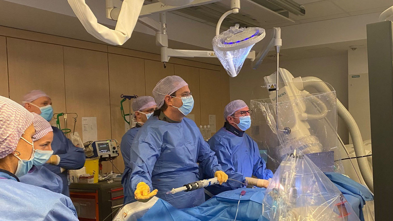
TAVI (Transcatheter Aortic Valve Implantation), also known as TAVR (Transcatheter Aortic Valve Replacement), is a minimally invasive method of replacing the aortic valve without traditional open-heart surgery. Instead of removing the damaged valve, doctors implant a biological prosthesis inside it, allowing the new valve to take over the function of regulating blood flow.
The procedure is usually performed through a small puncture in the groin. A catheter is inserted into the femoral artery and guided to the heart, where the new valve is positioned and expanded. In most cases, the procedure does not require opening the chest or using a heart-lung machine. It is often performed under local anesthesia with sedation.
This method is particularly suitable for older patients and those at increased risk of complications from open-heart surgery. In certain cases, TAVI can also be used to treat a previously implanted biological valve that is no longer functioning properly (a valve-in-valve procedure).
The main advantages of TAVI include less surgical trauma, a shorter hospital stay, and faster recovery compared with traditional surgical valve replacement. However, the procedure is not suitable for every patient.
SAVR — surgical aortic valve replacement
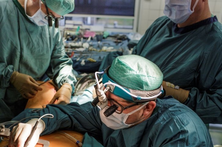
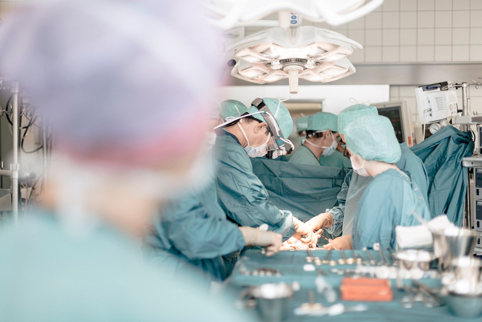
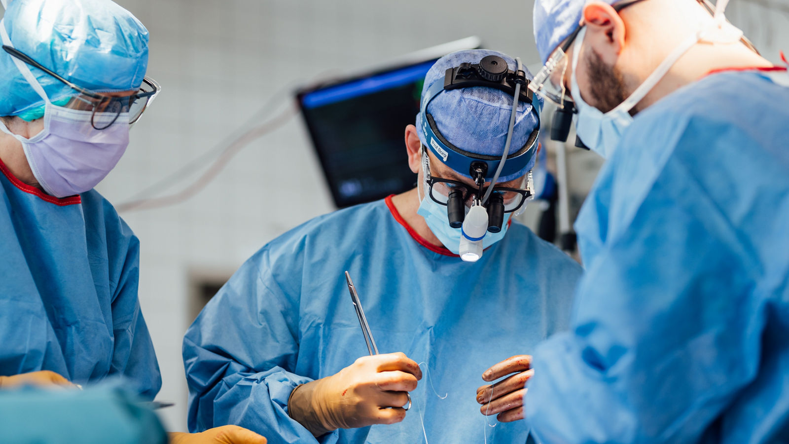
SAVR (Surgical Aortic Valve Replacement) is a traditional surgical procedure in which the damaged aortic valve is removed and replaced with a biological or mechanical prosthesis. Unlike TAVI/TAVR, the procedure is performed under general anesthesia using a heart-lung machine (cardiopulmonary bypass).
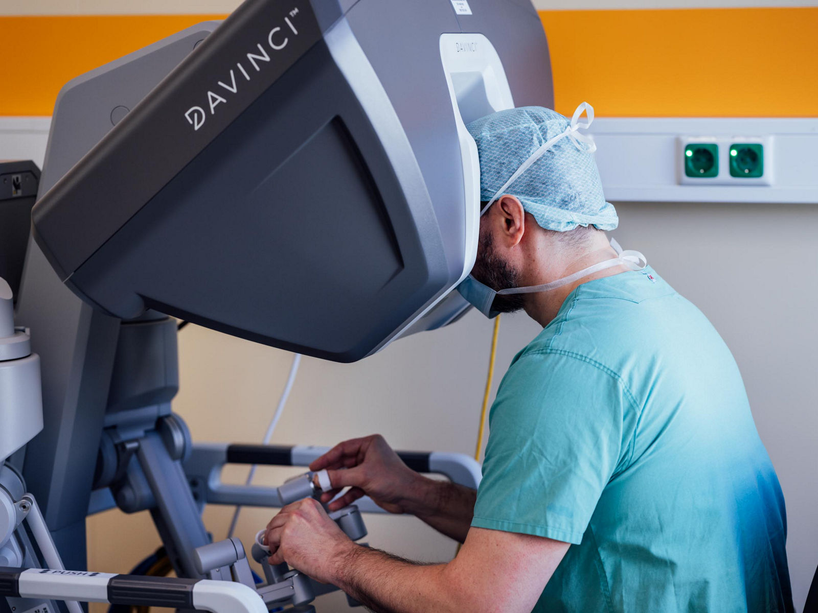
The operation is usually performed through an incision in the breastbone (sternotomy). However, in some cases, a minimally invasive approach through a smaller chest incision or partial sternotomy may be possible. After the patient is connected to the heart-lung machine, the surgeon removes the damaged valve, implants the prosthesis, and checks its function.
SAVR is particularly common in younger patients for whom valve durability is an important consideration, as well as in cases where TAVI is unsuitable due to anatomical characteristics or underlying medical conditions.
The main advantages of SAVR include the ability to implant either a mechanical or biological valve, directly remove damaged tissue, and perform additional cardiac procedures if necessary. However, compared with TAVI, SAVR is more invasive, requires general anesthesia, and typically involves a longer hospital stay and recovery period.
Ross procedure
The Ross procedure is a surgical method of aortic valve replacement in which the damaged aortic valve is replaced with the patient's own pulmonary valve. A donor valve (homograft) is then implanted in the pulmonary position. As a result, the procedure involves surgery on two heart valves.
The procedure is performed under general anesthesia using a heart-lung machine. The surgeon removes the diseased aortic valve and replaces it with the patient's pulmonary valve, along with a section of the surrounding blood vessel. The right ventricular outflow tract is then reconstructed using a donor valve graft.
The Ross procedure is primarily performed in children, adolescents, and young adults. It is particularly considered when avoiding a mechanical prosthesis and the associated need for lifelong anticoagulant therapy is important.
The main advantage of this method is the use of the patient's own living valve, which provides near-natural blood flow characteristics and, in children, can grow with the body. In addition, patients generally do not require lifelong anticoagulant therapy solely because of the procedure. However, the operation is technically complex, requires highly experienced surgeons, and carries a risk of dysfunction of both the transplanted valve and the prosthesis in the pulmonary position. Further interventions may be necessary over time, which is why the Ross procedure is primarily performed at specialized cardiac surgery centers.
Choosing the treatment method and preparing for surgery
The choice of aortic valve replacement or repair depends on the type of valve disease, the condition of the valve, and the patient's overall health. In German cardiac surgery centers, treatment decisions are usually made by a multidisciplinary team of specialists, including cardiologists, cardiac surgeons, and cardiac imaging specialists (Heart Team).
When selecting a treatment, doctors consider the patient's age, the severity of aortic stenosis or regurgitation, the condition of the valve leaflets, aortic anatomy, underlying medical conditions, and surgical risks. For example, transcatheter aortic valve implantation (TAVI) may be preferred for older patients with suitable anatomy, while surgical valve replacement or repair is more often considered for younger patients and those who require additional procedures on the heart or aorta. The expected durability of the valve and the need for anticoagulant therapy are also taken into account.
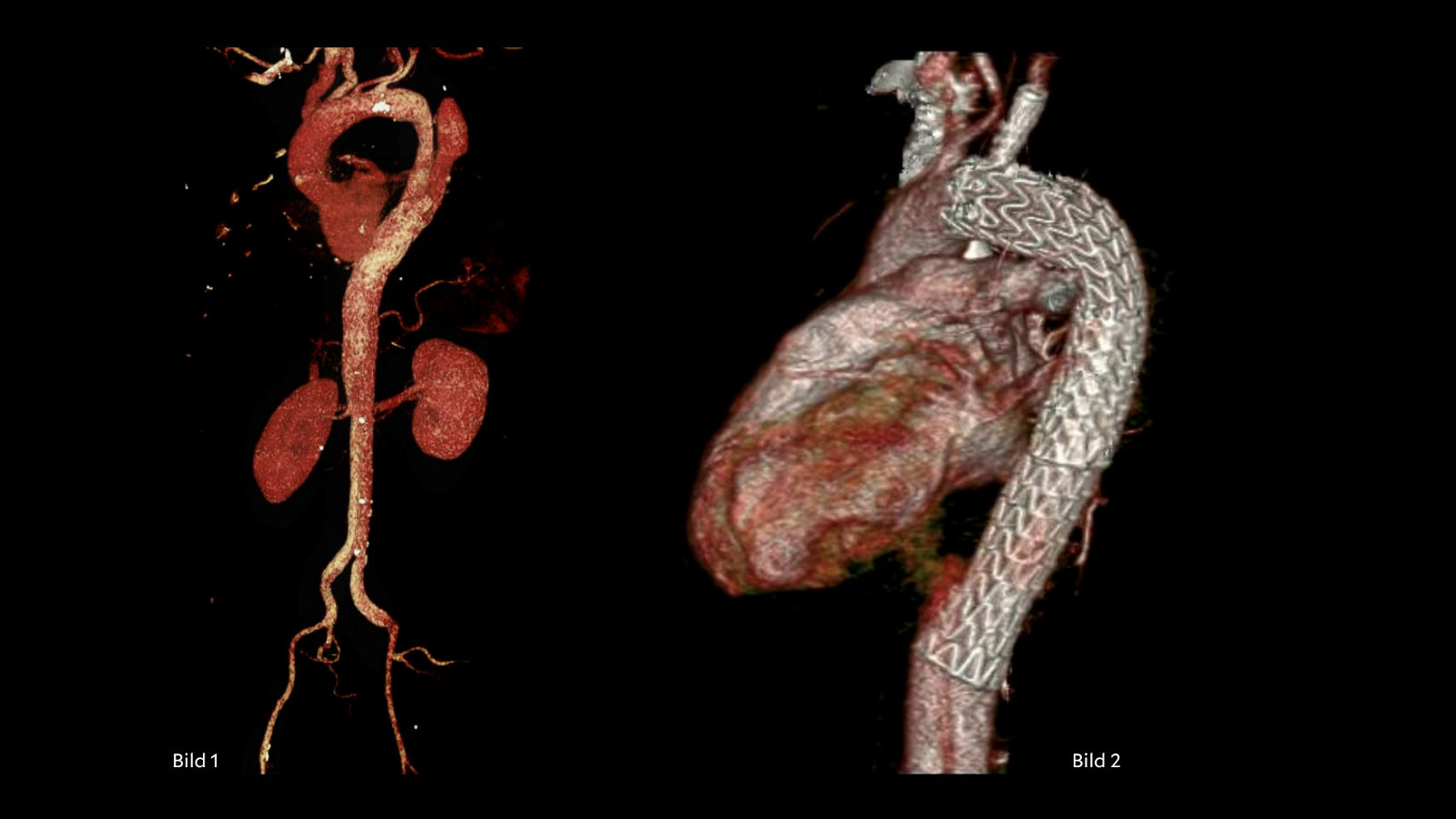
Before the procedure, the patient undergoes a series of examinations to assess heart function and determine the safest treatment approach. These typically include echocardiography, an electrocardiogram (ECG), and blood tests. Depending on the patient's condition, doctors may also recommend a CT scan of the heart and aorta, coronary angiography, or additional tests.
Patients should inform their medical team about all medications they take, allergies, and chronic health conditions. Particular attention is paid to anticoagulants and other medications that affect blood clotting, as any adjustments before surgery must be determined by the treating physician. It is also important to identify and treat active infections, including dental infections, before the procedure.
Before admission, patients receive individualized instructions regarding food and drink, medication use, and restrictions related to anesthesia. The preparation process varies depending on the selected treatment method, as open-heart surgery and transcatheter valve implantation may require different examinations and precautions.
Rehabilitation and life after surgery
Recovery after aortic valve replacement or repair depends on the type of procedure, the patient's age, and their overall health. After transcatheter aortic valve implantation (TAVI), patients generally recover more quickly and may be discharged from the hospital within a few days. Open-heart surgery usually requires a longer hospital stay and rehabilitation period, with full recovery taking several weeks or months.
In Germany, patients are often advised to undergo specialized cardiac rehabilitation (Kardiologische Rehabilitation) after heart surgery. The program may include therapeutic exercises, gradually increasing aerobic activity, breathing exercises, monitoring of heart rhythm and blood pressure, and guidance on nutrition and lifestyle. The aim of rehabilitation is to restore physical endurance, reduce the risk of complications, and help patients safely return to their daily activities.
Life after aortic valve replacement or repair
Following a successful procedure, many patients experience less shortness of breath, fatigue, and other symptoms, along with improved exercise tolerance. However, regular follow-up with a cardiologist remains essential regardless of the treatment method.
Long-term care depends on the type of valve implanted or the repair technique used:
- Mechanical valve — lifelong anticoagulant therapy, usually with vitamin K antagonists, is required, along with regular monitoring of the international normalized ratio (INR).
- Biological valve or TAVI — lifelong anticoagulant therapy is generally not required solely because of the valve. However, the doctor may prescribe antithrombotic medication depending on other medical conditions and individual indications.
- Native valve repair — generally avoids the need for lifelong anticoagulant therapy solely because of the procedure, but requires regular monitoring of the valve leaflets and valve function.
After discharge, patients should take their prescribed medications, gradually resume physical activity, and attend follow-up examinations, particularly echocardiography. Maintaining good oral hygiene and treating infections promptly are also important, as certain valve procedures are associated with an increased risk of infective endocarditis.
Most patients can return to their usual activities after successful treatment, including work, travel, and moderate exercise. The timing of recovery and the appropriate level of physical activity are determined individually, based on the type of procedure, heart function, and rehabilitation progress.
Aortic valve replacement and repair at Helios hospitals
Helios hospitals in Germany offer modern treatment options for aortic valve disease, ranging from minimally invasive transcatheter aortic valve implantation (TAVI) to surgical valve replacement and valve-sparing repair procedures.
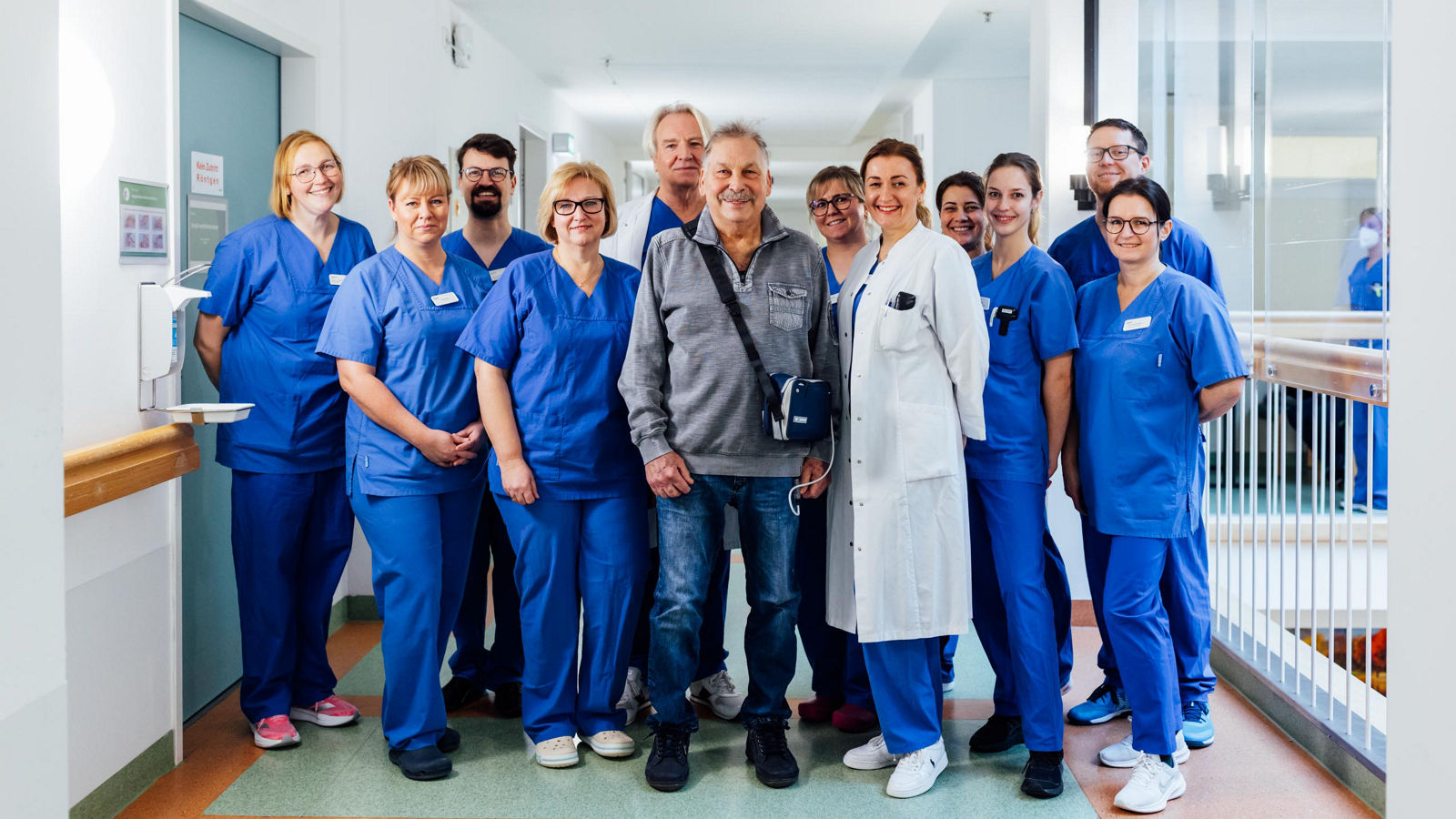
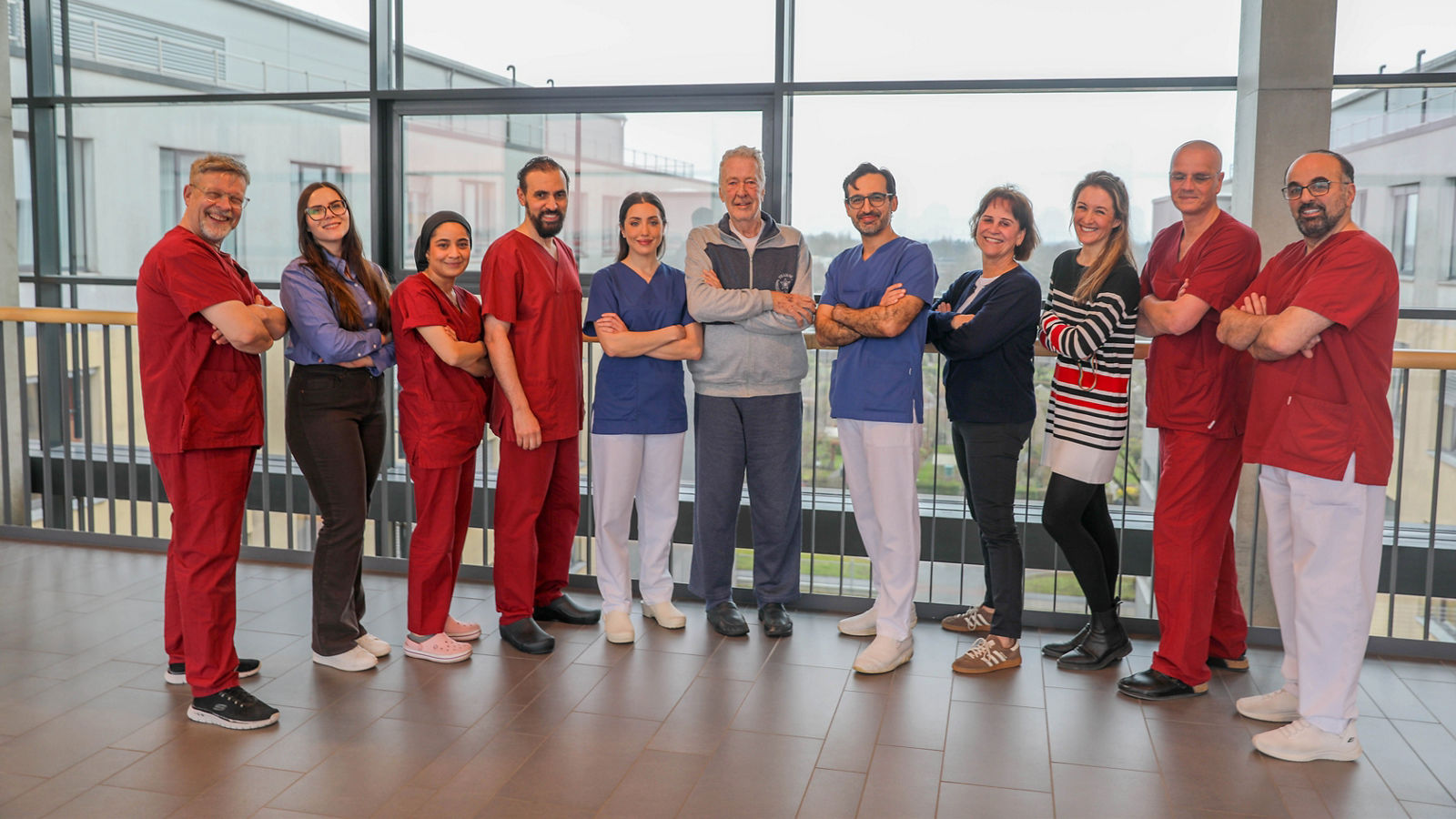
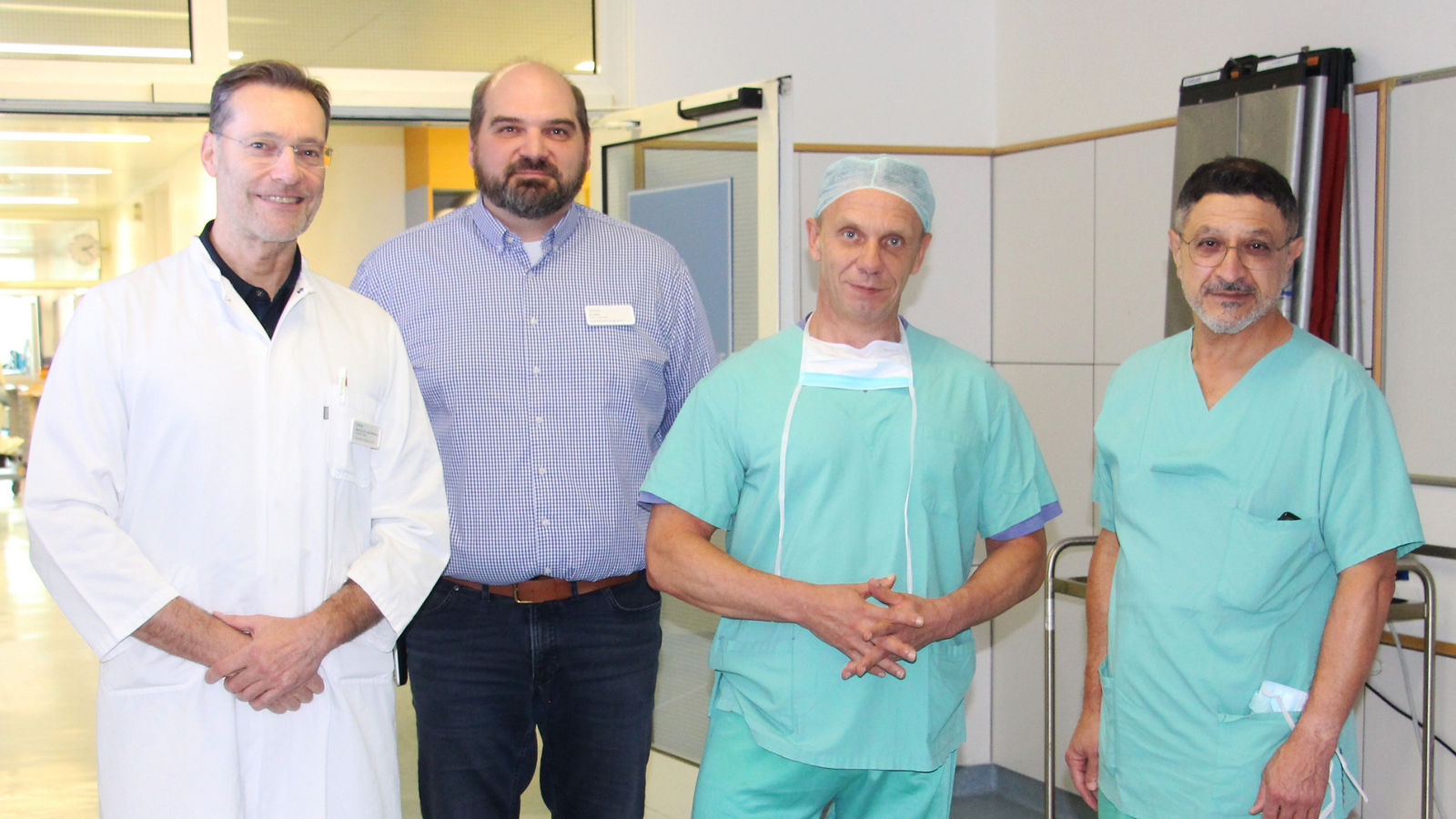
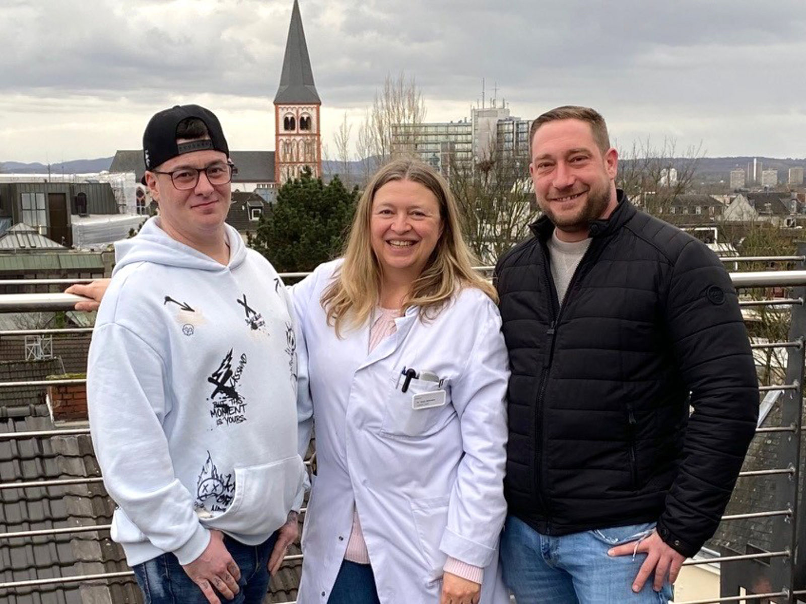
These procedures are performed at several Helios hospitals, including Helios Heart Center Leipzig, Helios Dr. Horst Schmidt Hospital in Wiesbaden, Helios Hospital Siegburg, and Helios Hospital Krefeld.
The treatment approach is tailored to each patient based on diagnostic findings, the condition of the valve, and their overall health. Specialists assess whether the patient's native valve can be repaired, whether replacement is necessary, and the potential risks of each procedure to determine the most appropriate treatment strategy.
If you are interested in arranging diagnosis or treatment at one of the Helios hospitals, you can submit a request using the contact form on our website or contact us via the email address provided at the top of the website.
Learn more about arranging treatment at Helios hospitals ⟶
Do you need more information about Helios Hospitals or want to schedule your treatment?